
1 Lab Practical 1
Introduction and Learning Objectives
Lab Practical 1 includes structures covered in the following chapters of your textbook:
-
- Chapter 1-A First Look at Anatomy
- Chapter 2-The Cell: Basic Unit of Structure and Function
- Chapter 4-Tissues Level or Organization
- Chapter 5-The Integumentary System
Introduction
Welcome to A215 Lab!
Chances are that you are taking this course because you are pursuing a degree in the health sciences. This class will provide you with a good foundation upon which all other courses related to the human body will build. In this section, we will get our feet wet by learning common anatomical terminology. From there, we will review the building blocks of this course beginning with a review of cellular anatomy. Did you know that when cells combine to perform a common function, they form what is known as a tissue? There are four primary tissue types in the human body including epithelial tissue, muscle tissue, connective tissue, and nervous tissue. Once we have a good grasp of the tissue types we will learn about their characteristics under a light microscope.
From there, we will move on to our first organ system; the Integumentary System, which we can see on the surface of our body. We tend to care a lot about this system because it is visible to us and to everyone around us. Consider how much money we spend each year for the benefit of this system; for example, shampoo for our hair, nail polish for our nails, and wrinkle cream for our skin.
We hope that this introduction has stimulated your interest in anatomy and you are ready to get started with the semester!
Pre-Test Assessment
Please complete the Pre-Test assessment for the lab material. This assessment does not count toward your course grade, but will provide us with an indication of your learning over the course of the semester.
Lab Practical Pre-Test Assessment; Does Not Count Toward Overall Course Grade
Course Learning Outcomes (CLO)
Identify and Understand Most Major Body Systems
Assessments used:
- Quizzes in the form of self-check assessments (formative)
- Lab Practical Exam (summative)
Module Learning Outcomes
Successful completion of this module demonstrates that you have met the following Module Level Outcomes (MLO). The numbers at the end of each MLO corresponds to the assessment used to measure attainment of the Course Learning Outcome (CLO).
Cell Model:
- Identify the organelles on the cell model (2)
- Understand the functions of the organelles on the cell model (1)
Mitosis Model:
- Identify the stages of mitosis on the mitosis models (2)
- Understand the specific characteristics of the phases of mitosis to include: prophase, metaphase, anaphase and telophase (1)
Cell Membrane Model:
- Identify the structures on the cell membrane model (2)
- Understand the characteristics of a phospholipid membrane to include the hydrophilic heads, hydrophobic tails and integral (transmembrane) protein (1)
Integumentary System Model:
- Identify the epidermis, dermis and hypodermis (2)
- Identify the layers within the epidermis (2)
- Identify the epidermal derivatives (2)
- Identify the structures within the dermis (2)
- Identify the structures within the hypodermis (2)
- Understand the functions of the structures listed on the integumentary system model (1)
Additional Knowledge:
- Identify all four tissue types (epithelial tissue, muscle tissue, connective tissue and nervous tissue) by using a light microscope (2)
- Identify the common locations for the tissue types in the human body (2)
- Identify the abdominopelvic quadrants in the human body (2)
First: Watch Dr. Gannon’s Videos
- The videos will introduce you to the models and, associated structures, that you are expected to learn for Lab Practical 1.
- You are encouraged to watch the videos multiple times as part of your preparation for Lab Practical 1.
Playlists
Cell Structures
Integumentary System Playlist
Second: Lab Practical 1 Review Slides
Review the document linked below. Be able to identify every structure that is labelled in the document.
- Hint: Learn the material by hand drawing each model, illustration and histology slide and adding the labels for each on your drawing.
Third: Quick Check Assessment (Quiz)-Formative Assessment
- First, watch the videos linked at the top of each assessment.
- Then, take the assessments in the form of a quiz. The quizzes can be taken multiple times to help you prepare for the lab practical.
- The Cell
- Mitosis
- Cell Membrane Model
- Integumentary System Model
Structures To Know-Lab Practical 1
- Rewrite the names of every structure on this list to help with retention, recall and spelling.
- Note: Structures may be found on multiple models, illustrations and histology slides.
Lab Practical 1 Structure List
Quadrants-Be able to identify and name the four abdominopelvic quadrants. You should also be able to identify the organs located within these quadrants.
Cell Membrane Model
- Hydrophilic Phosphate heads of the Phospholipid Membrane
- Hydrophobic Fatty Acid Tails of the Phospholipid Membrane
- Integral or Membrane Protein
Cell Model
- Vacuole
- Pinocytotic Vesicle
- Cytoplasm
- Smooth Endoplasmic Reticulum
- Nucleus
- Rough Endoplasmic Reticulum
- Lysosome
- Nucleolus
- Nuclear Pore
- Nucleoplasm
- Nuclear Envelope
- Mitochondria
- Cytoplasm
- Smooth Endoplasmic Reticulum
- Nucleus
- Rough Endoplasmic Reticulum
- Centrioles
- Centrosome
Mitosis Models-Know the following phases and their characteristics noted on Lab Practical Review PowerPoint.
- Interphase
- Early Prophase
- Late Prophase
- Metaphase
- Early Anaphase
- Anaphase
- Telophase
Tissues: Know the following histology slides and the structures/characteristics that are labelled on each slide. Also, where noted, know the typical locations for each tissue.
-
Epithelium
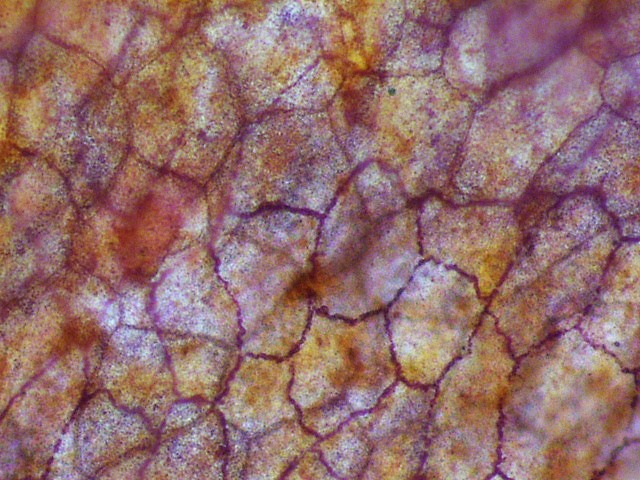
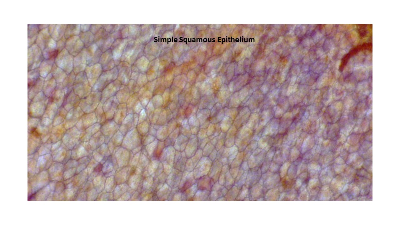
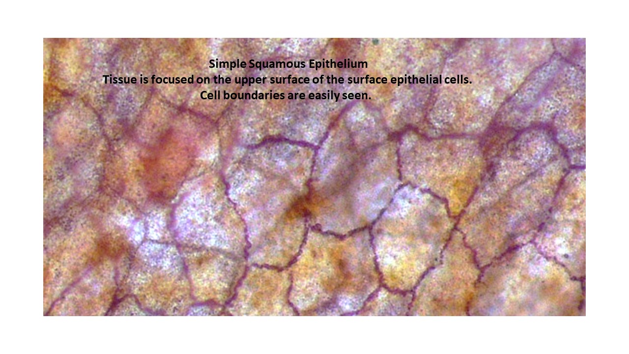
Simple Squamous Epithelium
- Simple Squamous Epithelium
- Simple Squamous Epithelium-Low Magnification
- Simple Squamous Epithelium-High Magnification
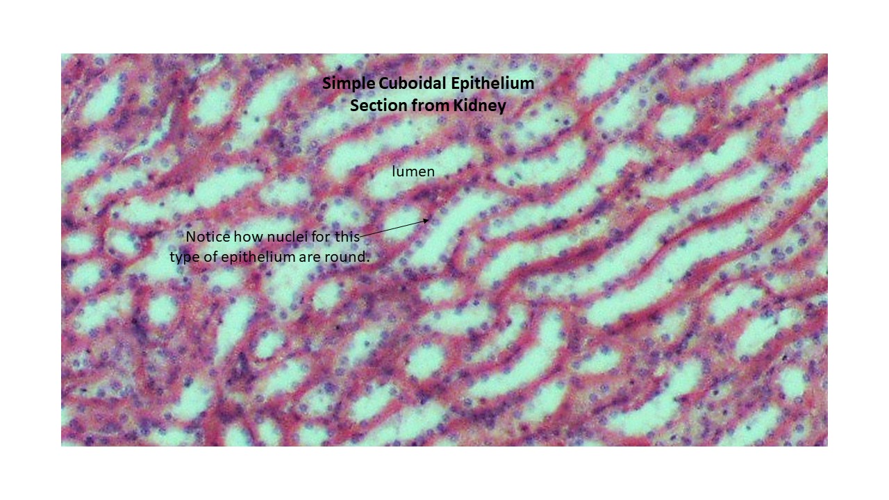
Simple Cuboidal Epithelium
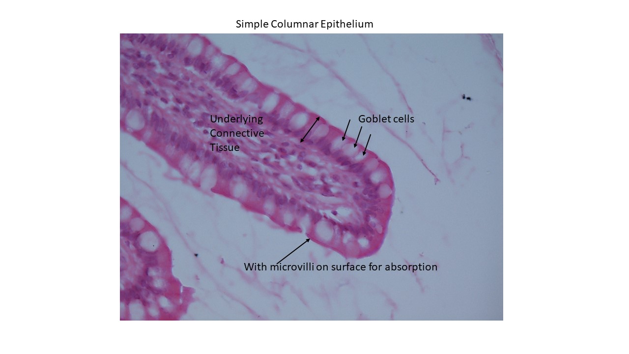
Simple Columnar Epithelium
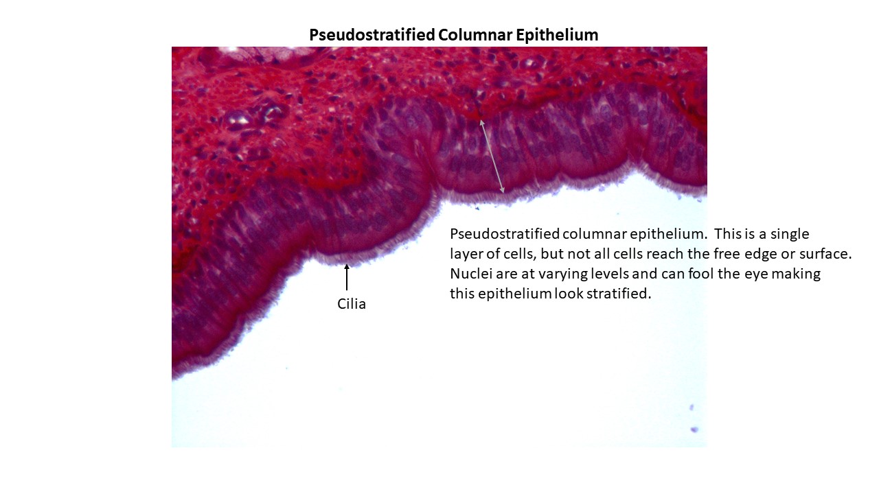
Pseudostratified Columnar Epithelium with Cilia
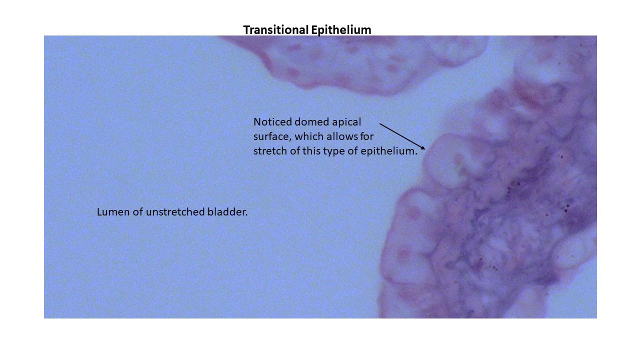
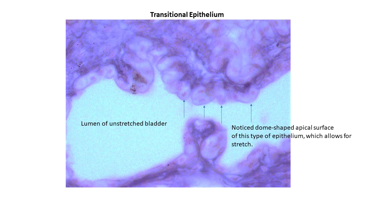
Transitional Epithelium
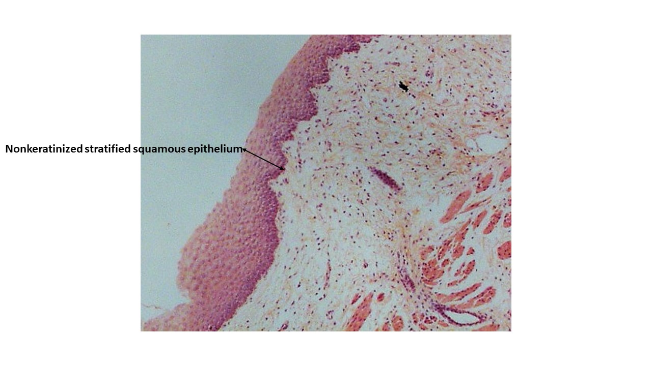
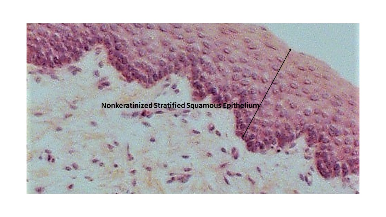
Nonkeratinized Stratified Squamous Epithelium
- Nonkeratinized Stratified Squamous Epithelium
- Nonkeratinized Stratified Squamous Epithelium-Higher Magnification
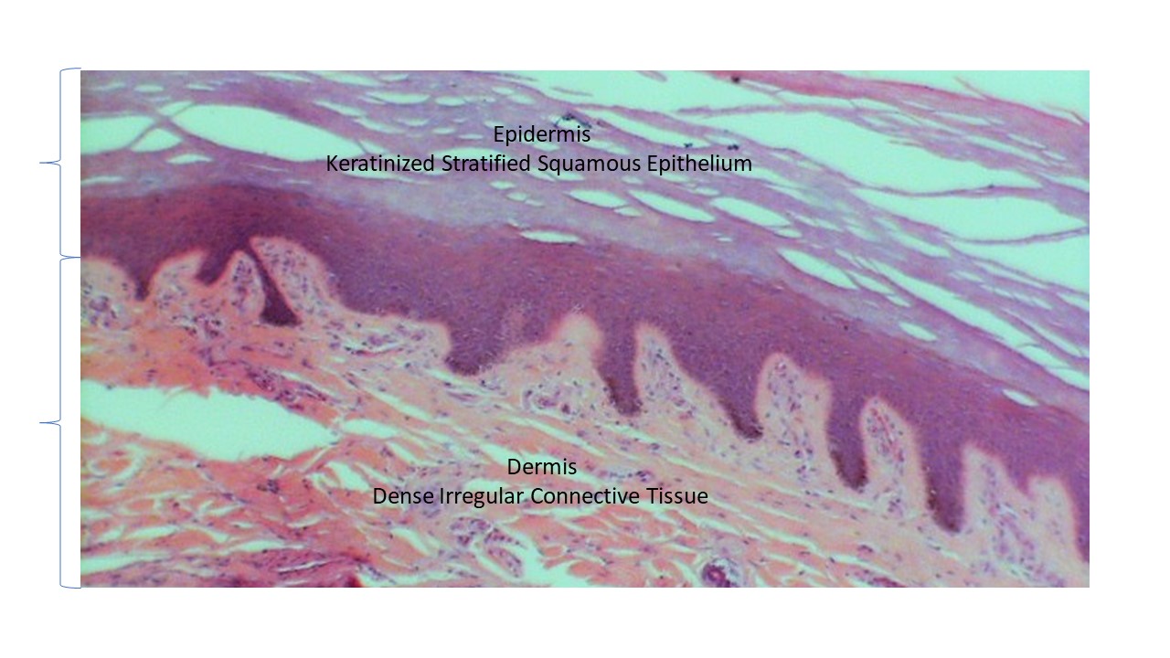
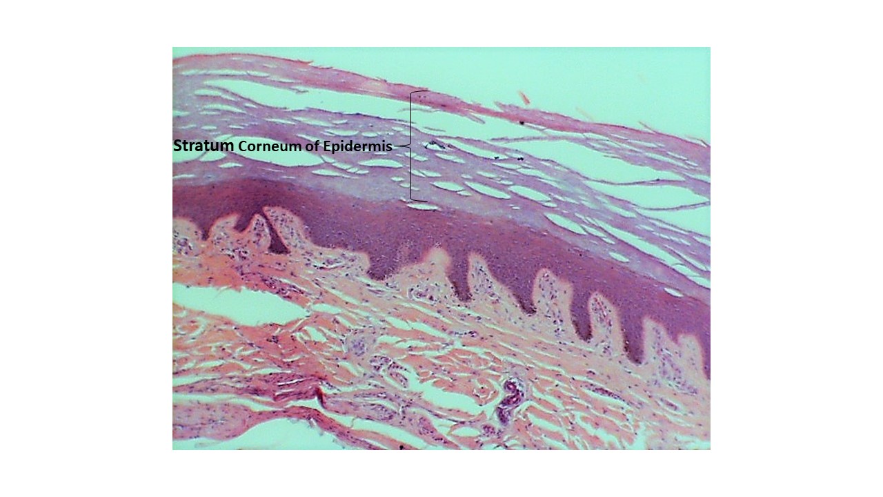
Keratinized Stratified Squamous Epithelium
- Keratinized Stratified Squamous Epithelium with Epidermis and Dermis
- Keratinized Stratified Squamous Epithelium with Stratum Corneum
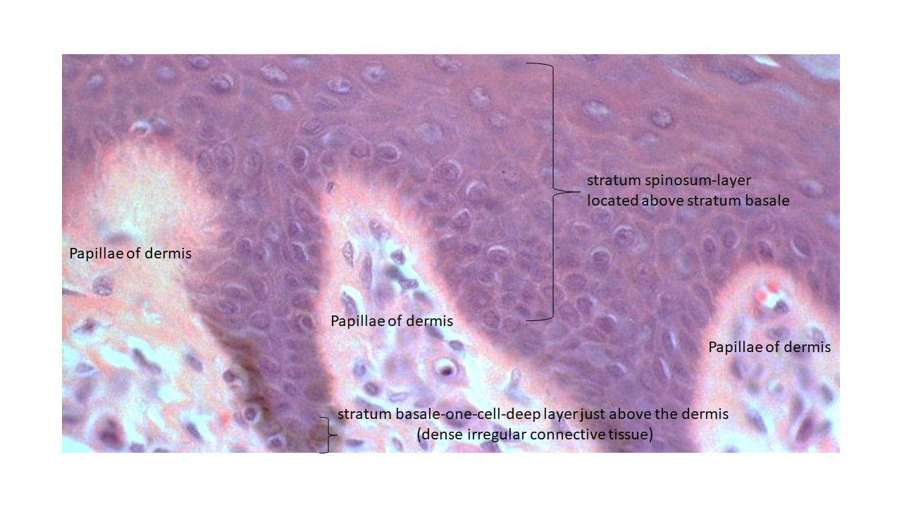
- Keratinized Stratified Squamous Epithelium with Stratum Spinosum, Stratum Basale and Dermal Papillae
- Keratinized Stratified Squamous Epithelium with Epidermis, Dermis and Hypodermis
Stratified Cuboidal Epithelium
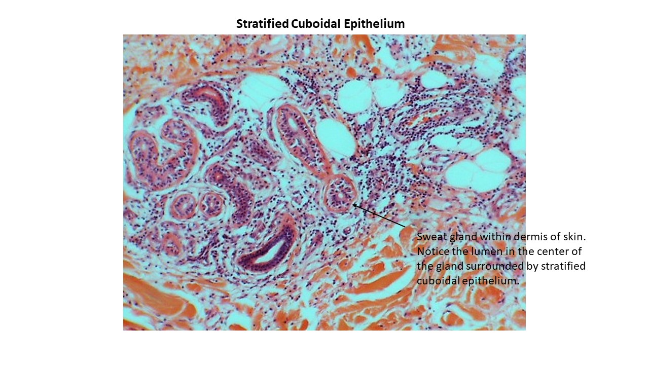
- Stratified Cuboidal Epithelium-Sweat Glands in Dermis of Skin
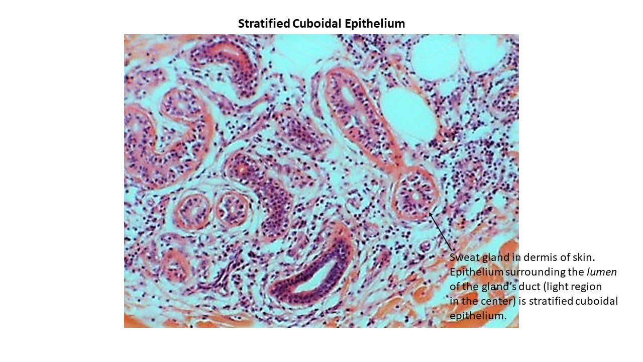
- Stratified Cuboidal Epithelium-Higher Magnification
Connective Tissue
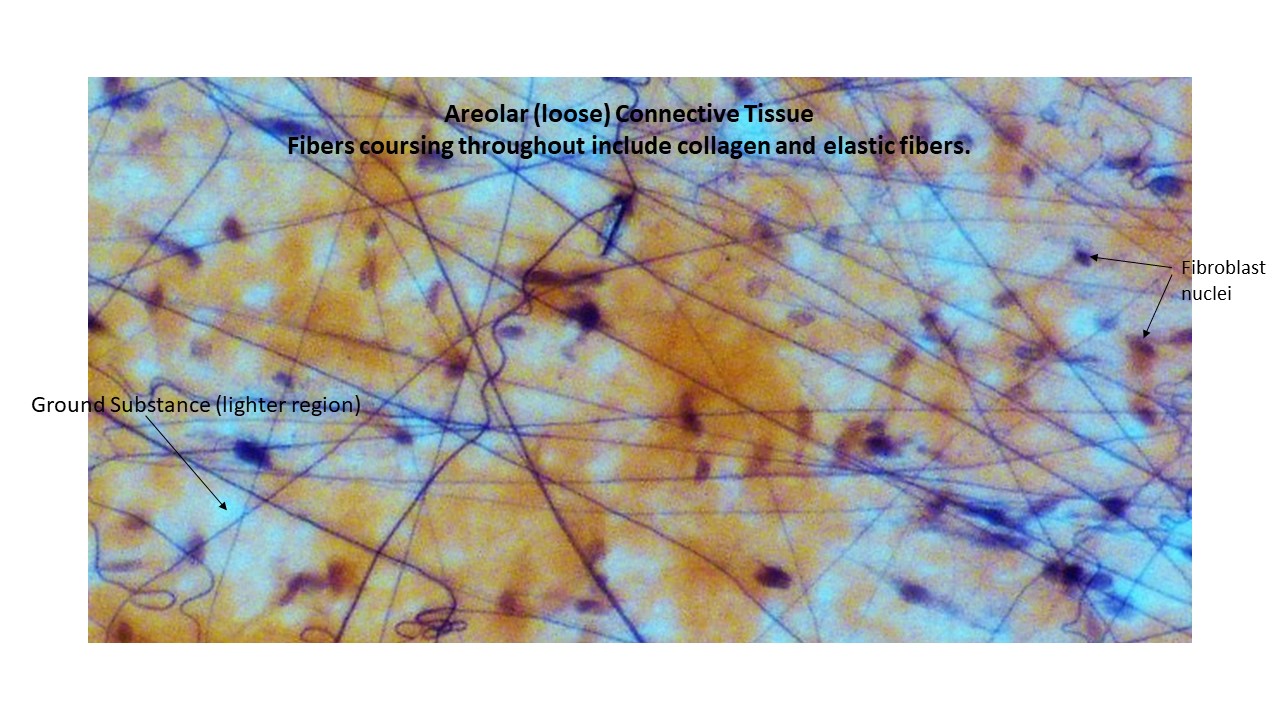
Areolar Connective Tissue
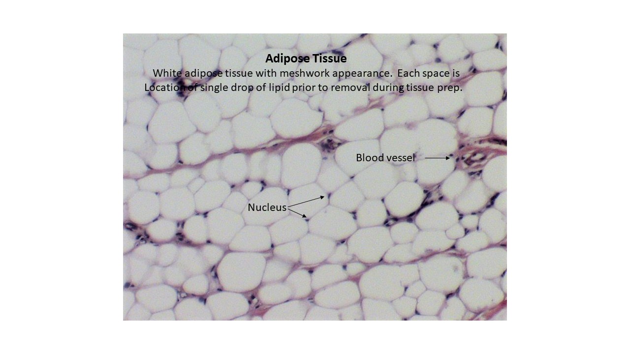
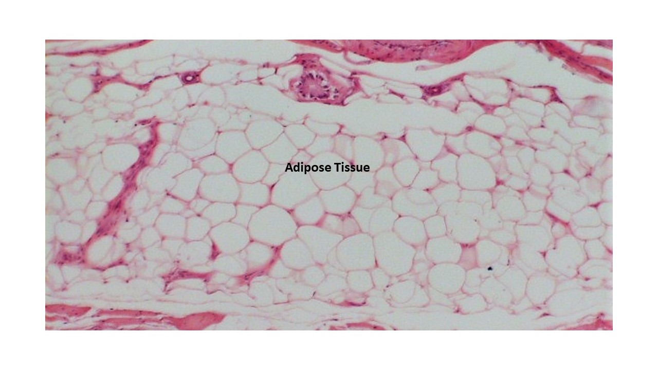
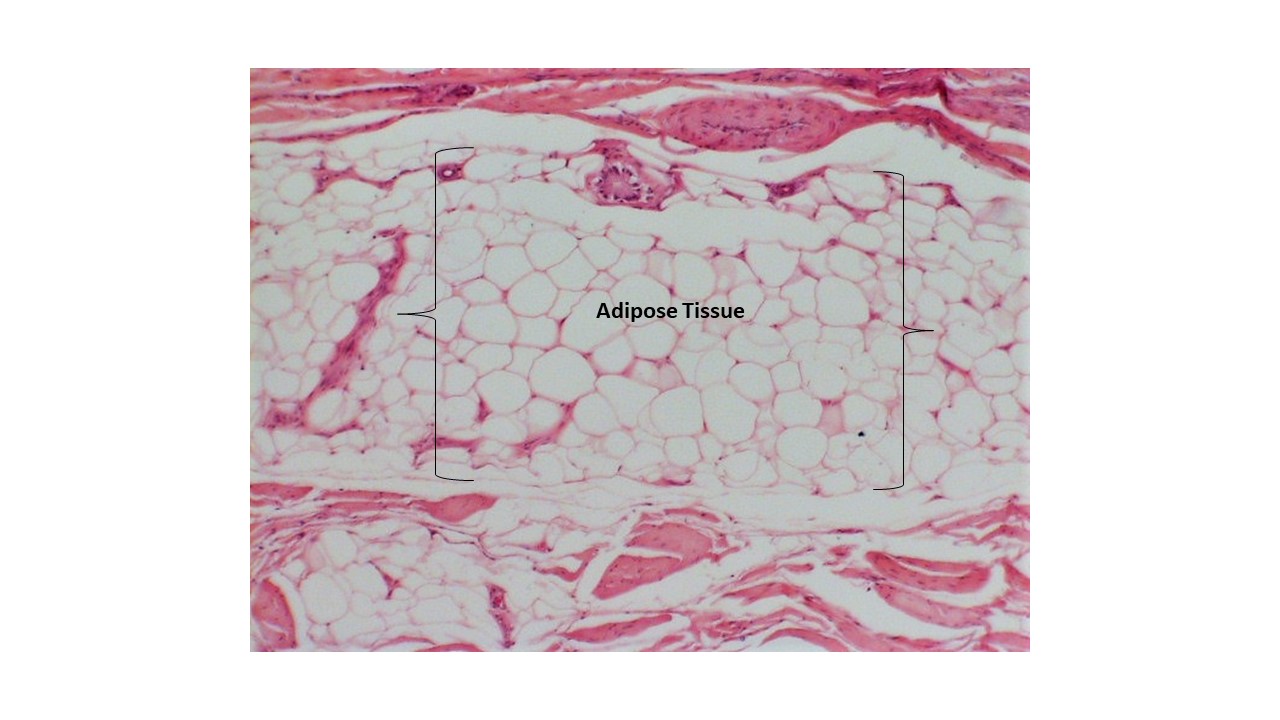
Adipose Connective Tissue
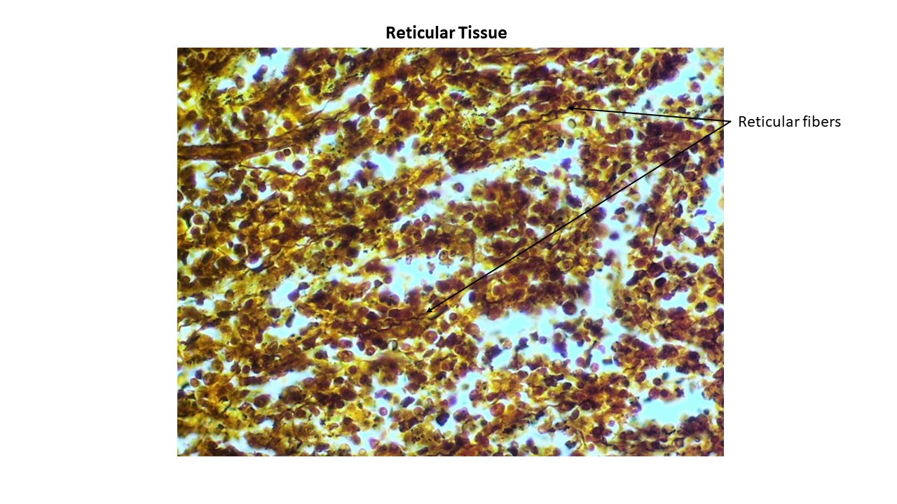
Reticular Connective Tissue
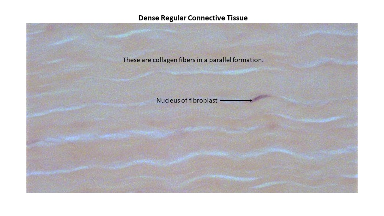
Dense Regular Connective Tissue
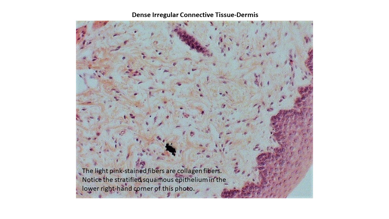
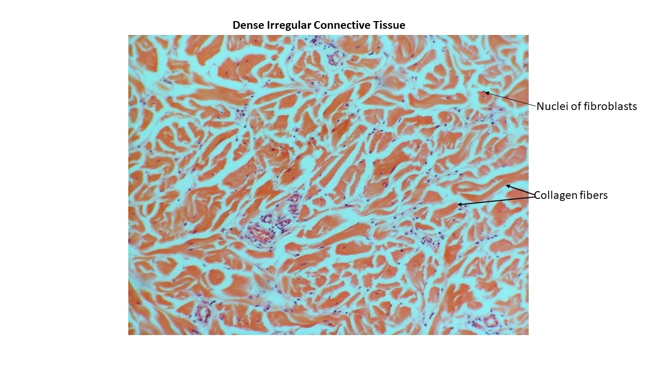
Dense Irregular Connective Tissue
- Dense Irregular Connective Tissue-Light Pink Stained Collagen Fibers
- Dense Irregular Connective Tissue-With Collagen Fibers and Fibroblasts
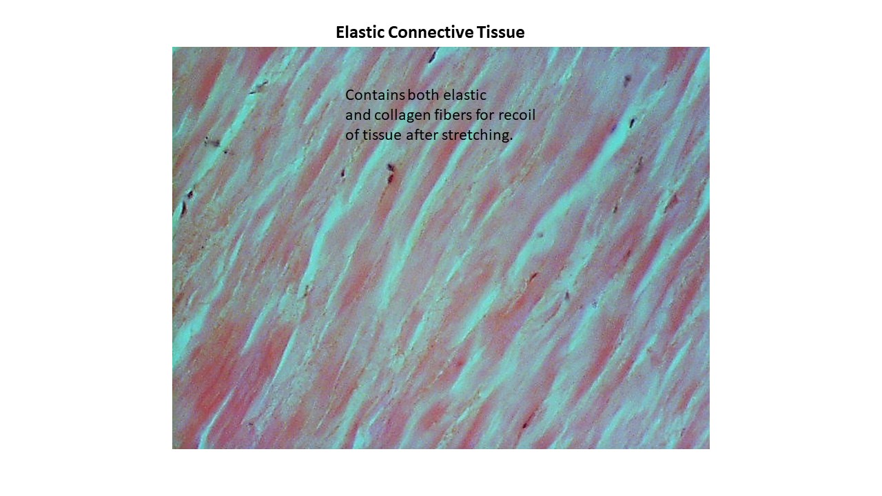
Elastic Connective Tissue
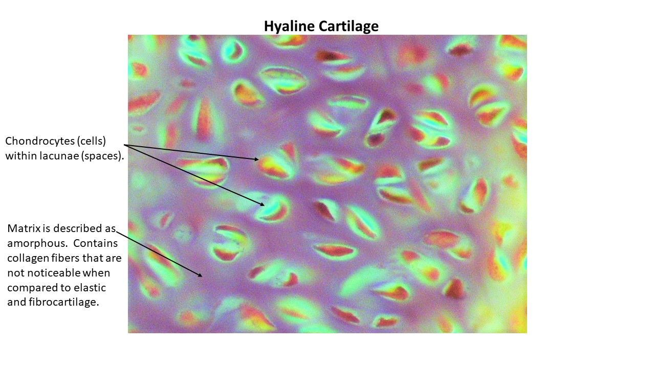
Hyaline Cartilage
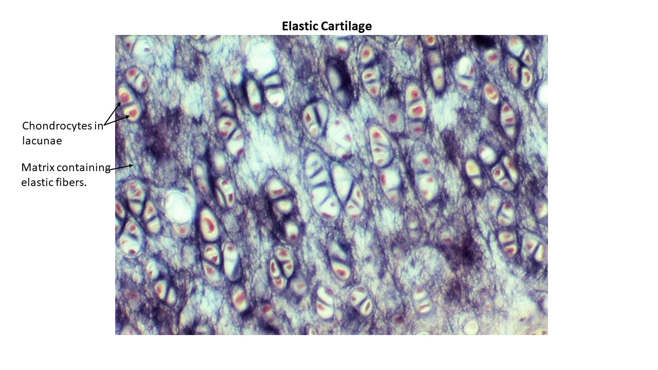
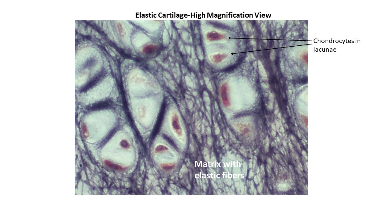
Elastic Cartilage
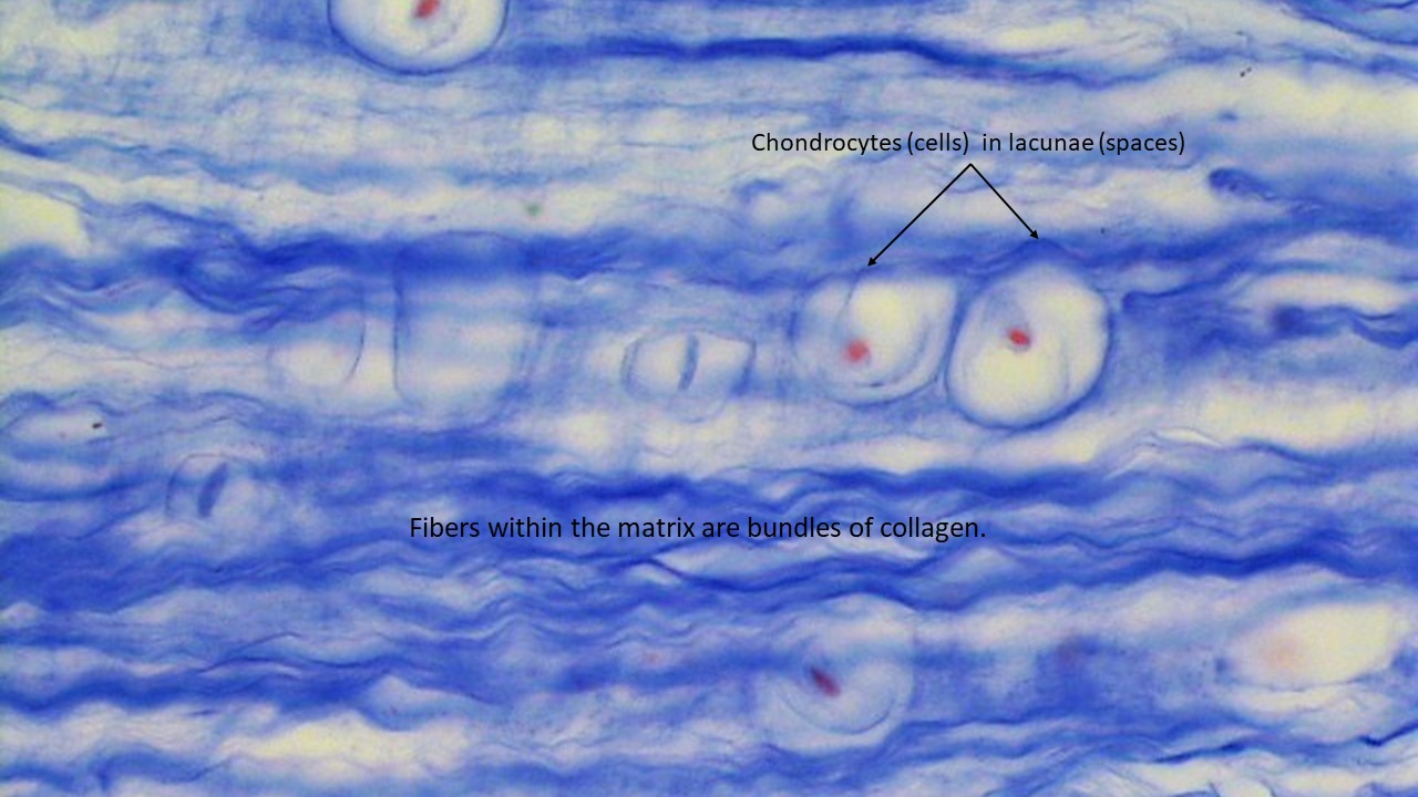
Fibrocartilage
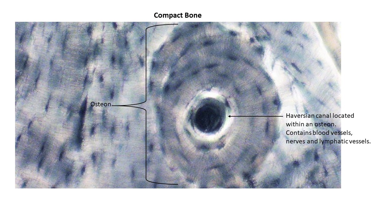
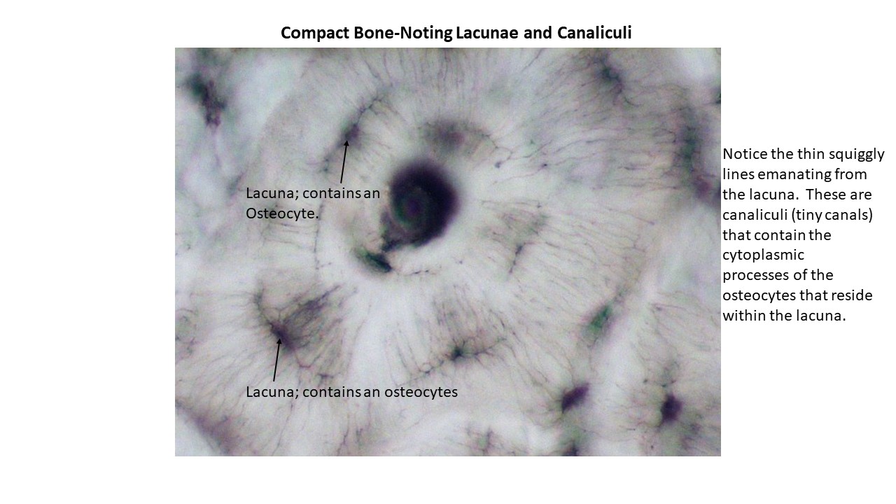
Bone (Compact)
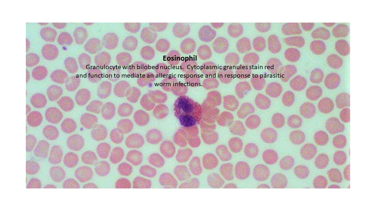
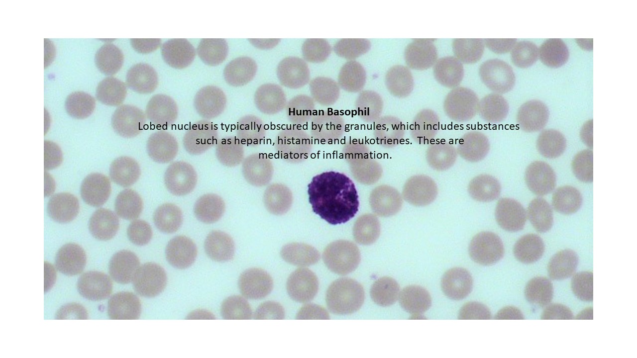
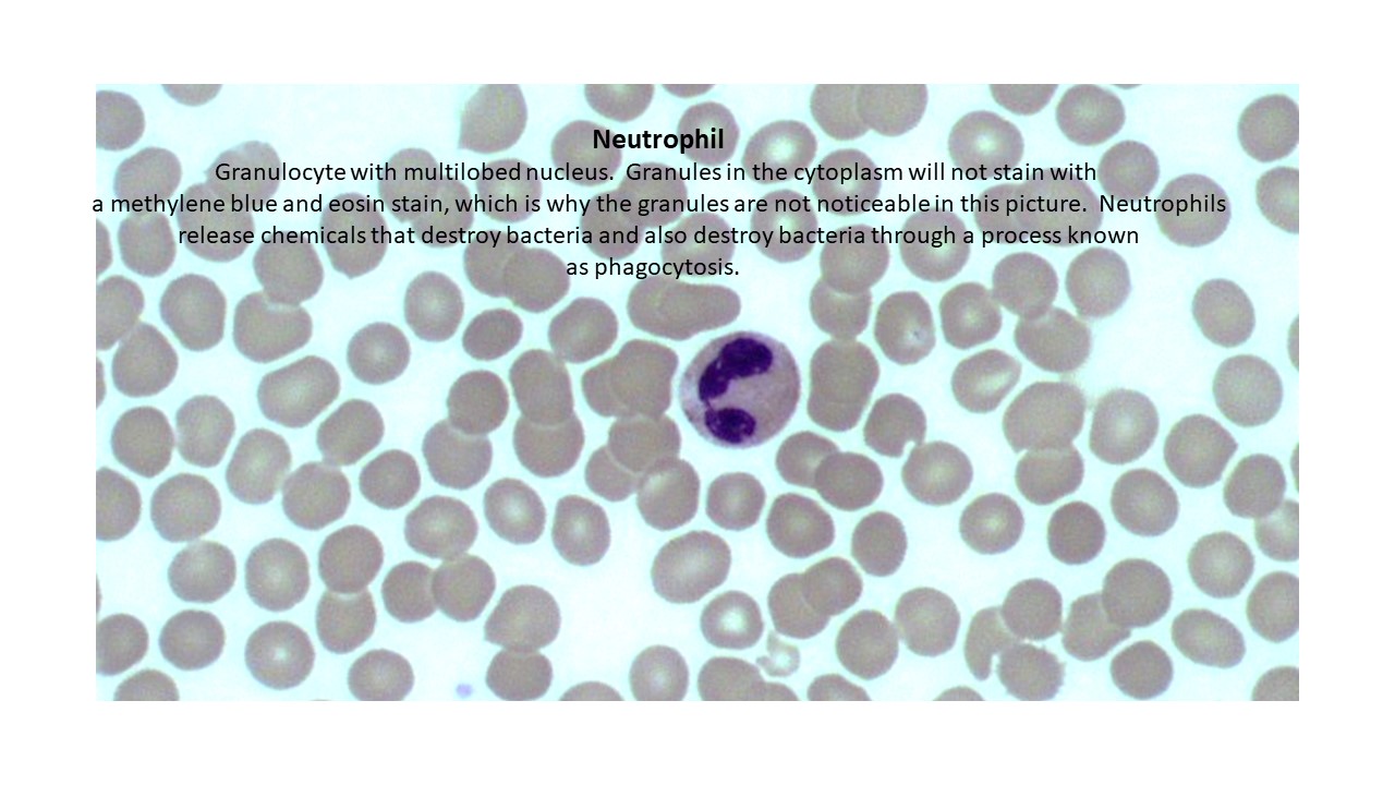
Blood-Granulocytes
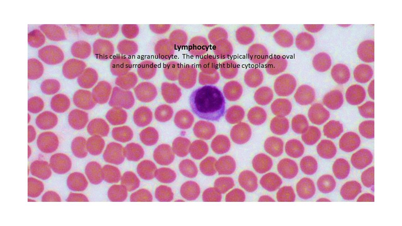
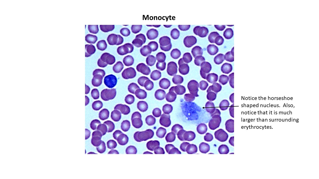
Blood-Agranulocytes
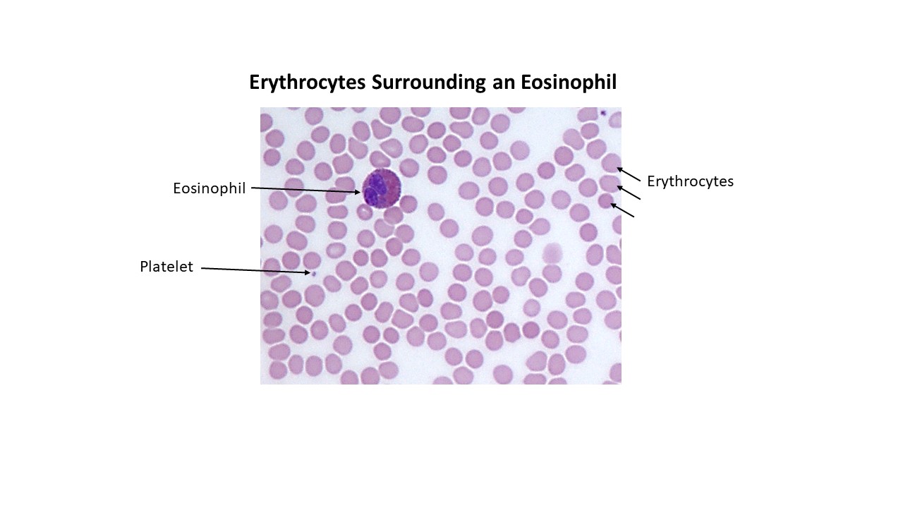
Blood-Erythrocytes and Platelet
Muscle Tissue
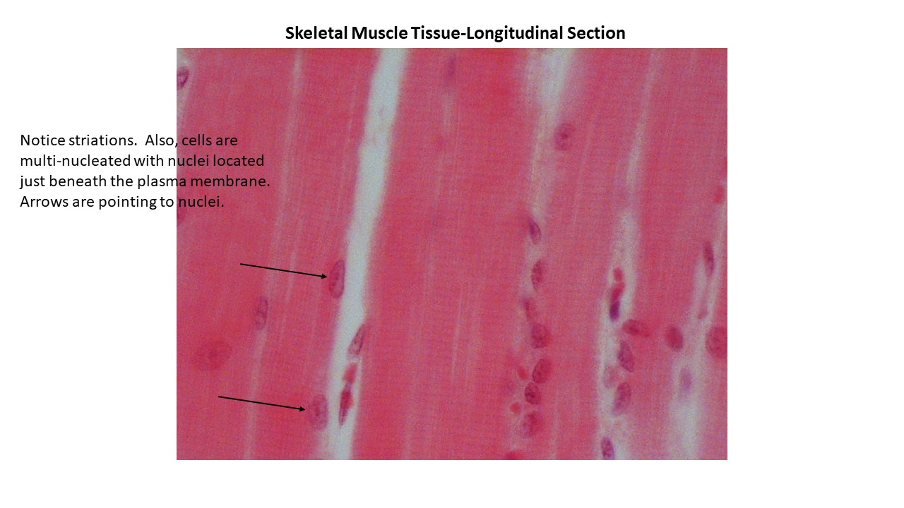
Skeletal Muscle
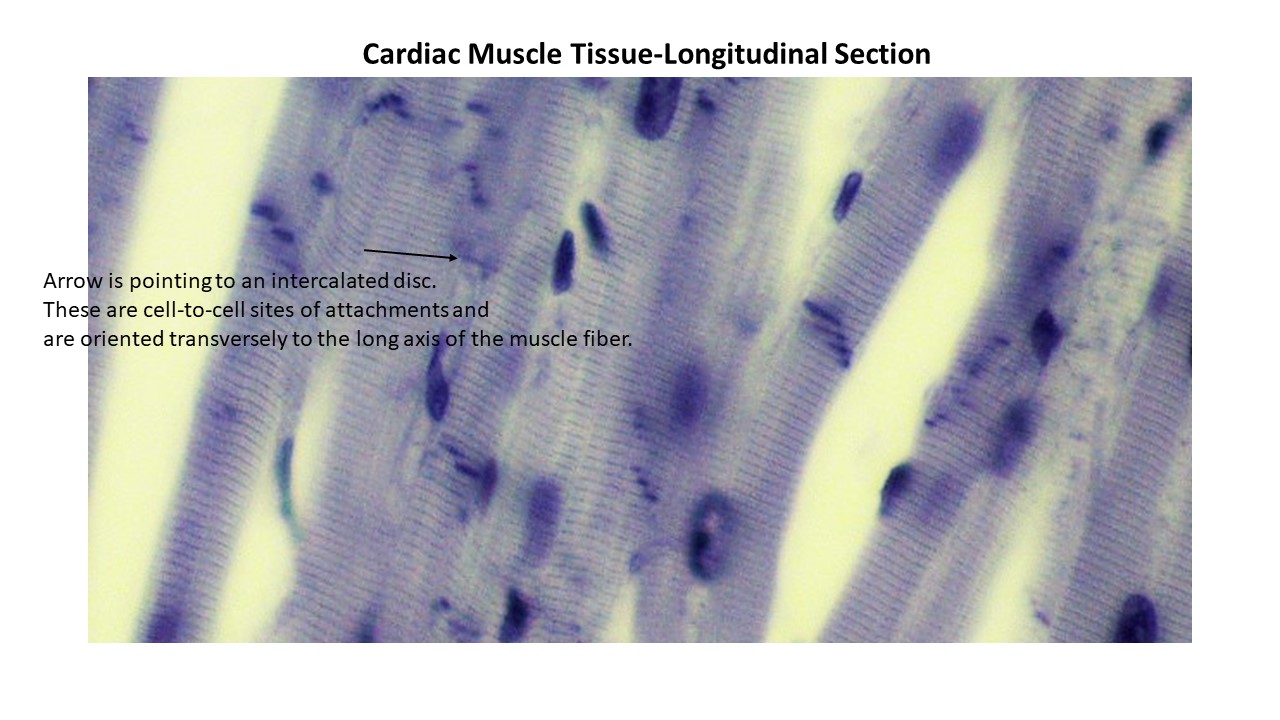
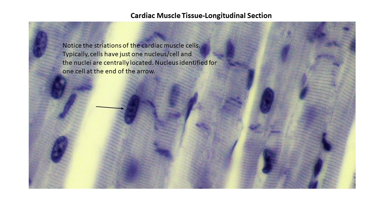
Cardiac Muscle
- Cardiac Muscle Tissue-Includes an Intercalated Disc
- Cardiac Muscle-Striations Evident and Nucleus Identified
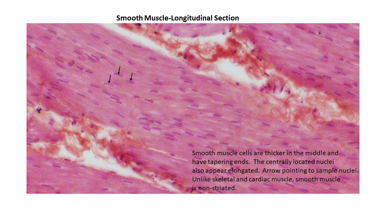
Smooth Muscle
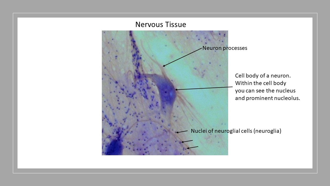
Nervous Tissue
Nervous Tissue
Integumentary Models
- Understand the difference between thin skin and thick skin
- Hair
- Hair Root Bulb
- Hair Root Plexus
Epidermis
- Stratum Basale
- Stratum Spinosum
- Stratum Granulosum
- Stratum Lucidum (thick skin only)
- Stratum Corneum
- Epidermal Ridges
Dermis-Dense Irregular Connective Tissue
- Dermal Papillae
- Arrector Pili Muscle-smooth muscle that contract involuntarily causing goosebumps.
- Pacinian Corpuscle
- Meissner’s Corpuscle
Hypodermis
- Adipose Tissue
Sweat Glands
- Merocrine Sweat Gland
- Apocrine Sweat Gland
- Sebaceous Gland