
3 Cleft Lip and Palate Repairs
Matt Hamilton, DO
Patients
The incidence is approximately 1:700 live births. Thought to have both environmental and genetic links. Cleft lip and palate are associated with over 400 syndromes. Of special anesthetic interest is the cleft palate associated with the Pierre-Robin sequence, which includes micrognathia (small jaw) and macroglossia (large tongue). Complete clefts may include extension into the nose, complete separation and protrusion of premaxilla, and complete clefting of the secondary palate. Secondary defects involve tooth development, nasal growth, and velopharyngeal function (swallowing and speech). Nearly all have middle ear disease due to eustachian tube abnormalities.
Surgery
The cleft lip surgery is usually performed around 3 months of age to help with maxilla formation and feeding. Cleft palate surgery is usually performed around 6-9 months of age and is vital to speech development. Alveolar clefts are closed, often using bone from rib, ilium, or skull. Pharyngeal flaps (posterior pharyngeal wall attached to the palate) are performed for velopharyngeal incompetence and are strongly associated with postoperative airway obstruction. Rhinoplasty and revisions are frequently performed. Ancillary procedures such as BMT’s or dental impressions are frequently performed at the same time as palate repairs.
Airway
Use a preformed cuffed oral RAE tube, the smallest cuffed ORAE is a 3.5, the smallest microcuffed oral RAE is a 3.0.
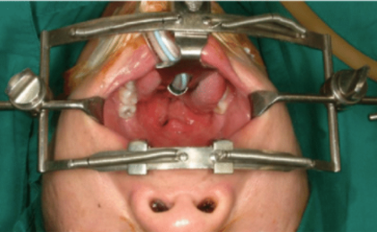
Secure oral RAE tube to the middle of the lower lip. Realize the face will be prepped with betadine so make sure it is secure. Try to provide a small amount of lateral mobility in the tube to allow the surgeon to place the tube in the groove of the tongue blade of the Dingman retractor. Observe ETT and ventilation pressures carefully after the surgeon positions the Dingman, which frequently compresses or kinks the ETT.
Inadvertent extubation is the single biggest hazard in these cases, so always have a spare (conventional) endotracheal tube available.
Inspissated secretions or blood is the second biggest hazard. Minimized with the use of cuffed ETT and PEEP.
Be sure humidifier working. Do not turn it off even if the patient is warm. Remember that 100% humidified gases at room temp (21 C) contain only 18 mg/L of water, vs 44 mg/L of water for gases at 37 C. Therefore, the relative humidity of gases that were fully saturated at room temp is only 40% saturated at body temp.
Intubation “tricks” include placing gauze in the cleft to keep your laryngoscope from sliding into the cleft, approaching the airway from the far right side of the mouth to give you enough room to maneuver the tube to intubate, and holding the RAE tube between your fingers in such a way that it straightens out at the acute bend while you are trying to pass it.
For cleft palate surgery make sure to have good IV access as the patient frequently refuse PO fluids.
Monitoring
Pulse oximetry, EtCO2, and peak airway pressures are key, since a dislodged, kinked or occluded ETT is the main intraoperative misadventure in these cases.
Temperature is monitored in the axilla to keep the probe out of the surgical field.
EXTUBATION: Extubate awake, moving all four extremities, eyes open. Use the 6 G’s. Be absolutely sure the throat pack has been removed. Blood and secretions in their mouths make these patients very susceptible to post-extubation laryngospasm. Reintubation is complicated by the presence of a fresh repair. Some patients with marginal airways will have a tongue stitch placed by the surgeon so that gentle traction can help keep the airway open in an emergency, although this is becoming less frequent. Other patients may have one or two nasal trumpets placed by surgery.
Pain Control
Cleft lip patients should have bilateral infraorbital blocks with 0.25% bupivacaine placed by surgery near the end of the procedure. They should wake up pain-free and usually require minimal narcotics in the PACU. Feeding is normally the answer to an upset child post cleft lip repair.
Cleft palate repair is far more uncomfortable. A multipronged approach is very useful. You should discuss a plan with your attending in advance. Preoperative oral Tylenol or a Tylenol suppository after induction. Ketorolac 0.5 mg/kg (max 15 mg) after the procedure is over. Morphine 50-100 mcg/kg prior to extubation. Dexmedetomidine 0.5-1 mcg/kg provides some sedation and analgesia without respiratory depression. The surgeon will use local with epinephrine which helps with pain and with hemostasis during the procedure. The APS is now performing V2 blocks.
Dental Impressions
For any cleft lip, cleft palate, or primary alveolar bone graft patient in whom dental impressions are being obtained as part of the procedure, tape the endotracheal tube in the midline of the lower lip.
References
Shapiro B. Humidity and aerosol therapy, in Clinical Applications of Respiratory Care. Year Book Medical Publishers. 1975; 165.
Mensil M, et al. A new approach for peri-operative analgesia of cleft palate repair in infants: the bilateral suprazygomatic maxillary nerve block. Paediatr Anaesth. 2010; 20: 343-349.
Woo J-M, Choi J-Y. Tonsillectomy as prevention and treatment of sleep-disordered breathing: A report of 23 cases. Maxillofacial Plastic and Reconstructive Surgery. 2016;38.
2/02 TWolfe
6/19 MHamilton