
3 Lab Practical 3
Introduction and Learning Objectives
Lab Practical 3 contains structures from the following chapters in your textbook
- Chapter 21-The Cardiovascular System: Blood
- Chapter 22-The Cardiovascular System: The Heart
- Chapter 23-The Cardiovascular System: Vessels and Circulation
Introduction
Many of us go about our daily lives without giving much consideration to the importance of the cardiovascular system. Did you know that your cardiovascular system works intimately with other organ systems? For example, think about your respiratory system. When you inhale, the oxygen that enters the alveoli of your lungs relies upon circulating blood to deliver that oxygen to the tissue cells of your body. Similarly, think about the last time you ate and how the nutrients that you consumed were delivered to your cells. As food is absorbed across the epithelial lining of your digestive system, it is your circulating blood that delivers these nutrients to the tissue cells. The heart, or the pump, that moves blood throughout your body must be healthy in order to deliver oxygen and nutrients where they are needed, and the blood vessels that deliver this blood will branch and distribute to ensure that your cells receive what they need.
It is likely that you or someone you know has a disorder that impacts the cardiovascular system. For example, sickle cell anemia is a disorder of the blood, cardiac arrhythmias and congestive heart failure affects our heart, and arteriosclerosis is an example of coronary artery disease. As we study the cardiovascular system in this section, take the time to consider these disorders and their potential impact on an individual’s daily life.
Now that you have an idea of what this section entails, let’s gets started with our study of the cardiovascular system.
Course Learning Outcomes (CLO)
Identify and Understand Most Major Body Systems
Assessments used:
- Quizzes in the form of self-check assessments (formative)
- Lab Practical Exam (summative)
Module Learning Outcomes
Blood Slides
- Identify an erythrocytes and platelet on a blood slide (2)
- Identify the various leukocytes using the key characteristics noted for each (2)
Heart Models
- Identify key anatomical structures on the external anatomy of the heart (2)
- Identify key anatomical structures on the internal anatomy of the heart (1) (2)
- Identify the major blood vessels that deliver blood to the heart and carry blood away from the heart (1) (2)
- Identify the coronary arteries and cardiac veins (1) (2)
Pig Heart (Dissected)
- Identify key anatomical structures on the external anatomy of the pig heart (2)
- Identify key anatomical structures on the internal anatomy of the heart (2)
- Identify the major blood vessels that deliver blood to the heart and carry blood away from the heart (2)
Blood Vessel Models
- Identify the blood vessels associated with the pulmonary circulation (1) (2)
- Identify the blood vessels associated with the systemic circulation (1) (2)
Blood Flow Through the Heart
- Understand the flow of blood through the human heart (1)
- Understand the flow of blood through the systemic arteries and veins (1)
First: Watch Dr. Gannon’s Videos and Dr. Zimmerman’s Dissection Videos
- The videos will introduce you to the models and, associated structures, that you are expected to learn for Lab Practical 3.
- You are encouraged to watch the videos multiple times as part of your preparation for Lab Practical 3.
Playlists
Circulatory System
Dissection Videos
- Surface Structures of the Heart
- Dissection of the Heart Part
- Identification of Interior Structures of the Heart
- Vessels that Service the Heart
Second: Lab Practical 3 Review Slides
Review the documents linked below. Be able to identify every structure that is labelled in the documents.
- Hint: Learn the material by hand drawing each model, illustration and histology slide and adding the labels for each on your drawing.
Virtual Lab Practical 3 Review Pictures_elab
Third: Quick Check Assessments (Formative Assessment)
- First, watch the videos linked at the top of each assessment.
- Then, take the assessments in the form of a quiz. The quizzes can be taken multiple times to help you prepare for the lab practical.
Structures To Know Lab Practical 3
- Rewrite the names of every structure on this list to help with retention, recall and spelling.
- Note: Structures may be found on multiple models, illustrations and histology slides.
The Cardiovascular System: Blood
Blood Histology Slides– Be able to identify the following blood cells and any key characteristics that are labelled.
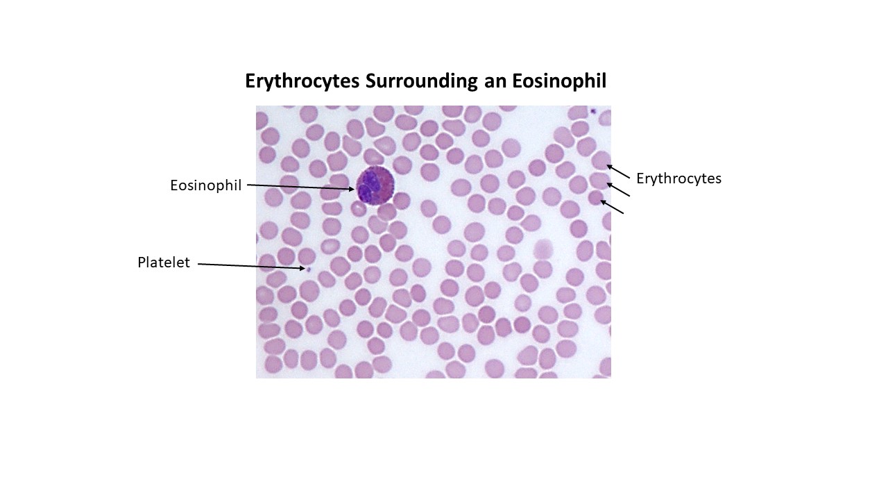
- Erythrocytes-Erythrocytes and Platelet
- Granulocytes
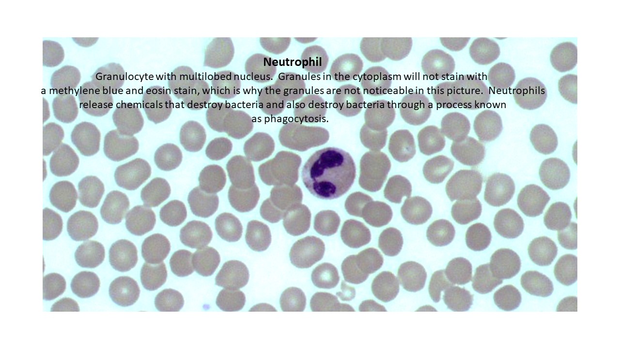
- Neutrophil-Neutrophil
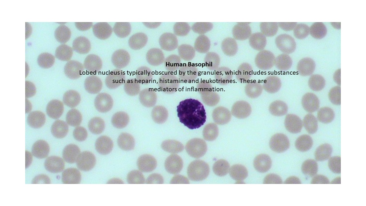
- Basophil-Basophil
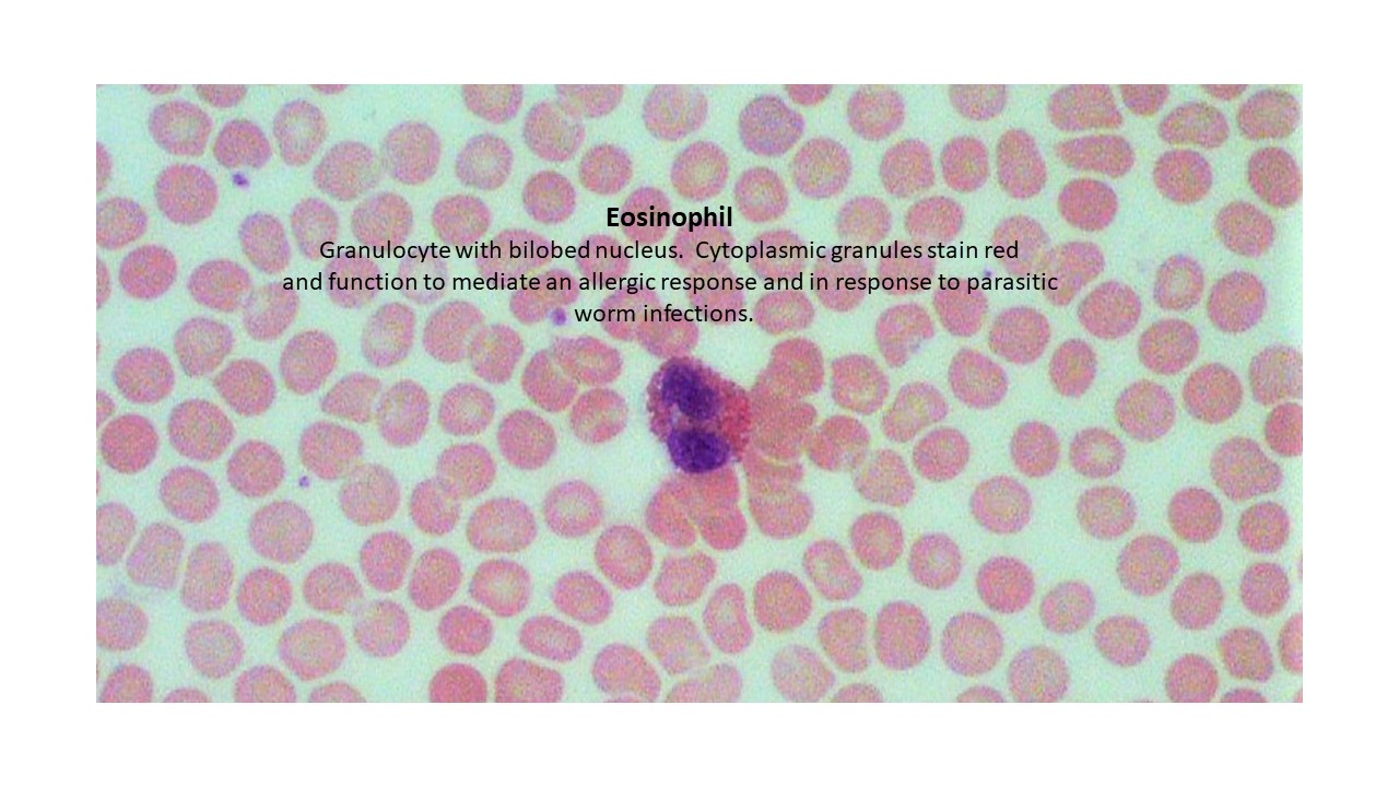
- Eosinophil-Eosinophil
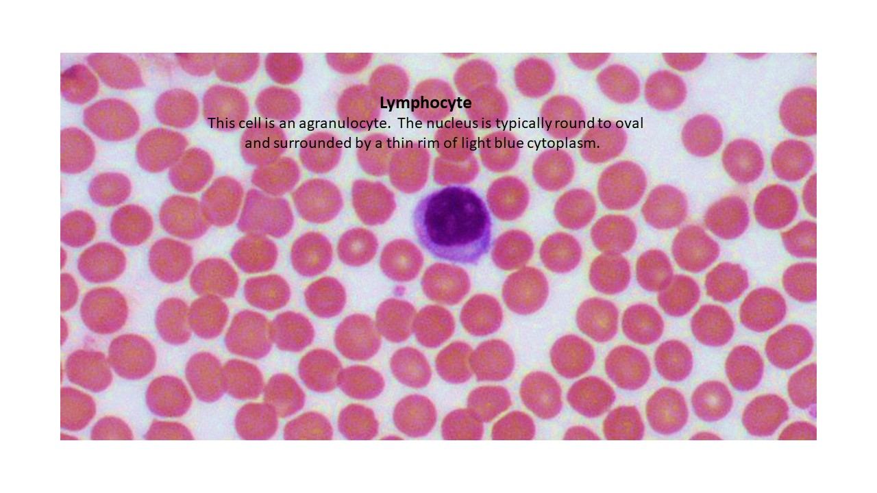
- Agranulocytes
- Lymphocyte-Lymphocyte
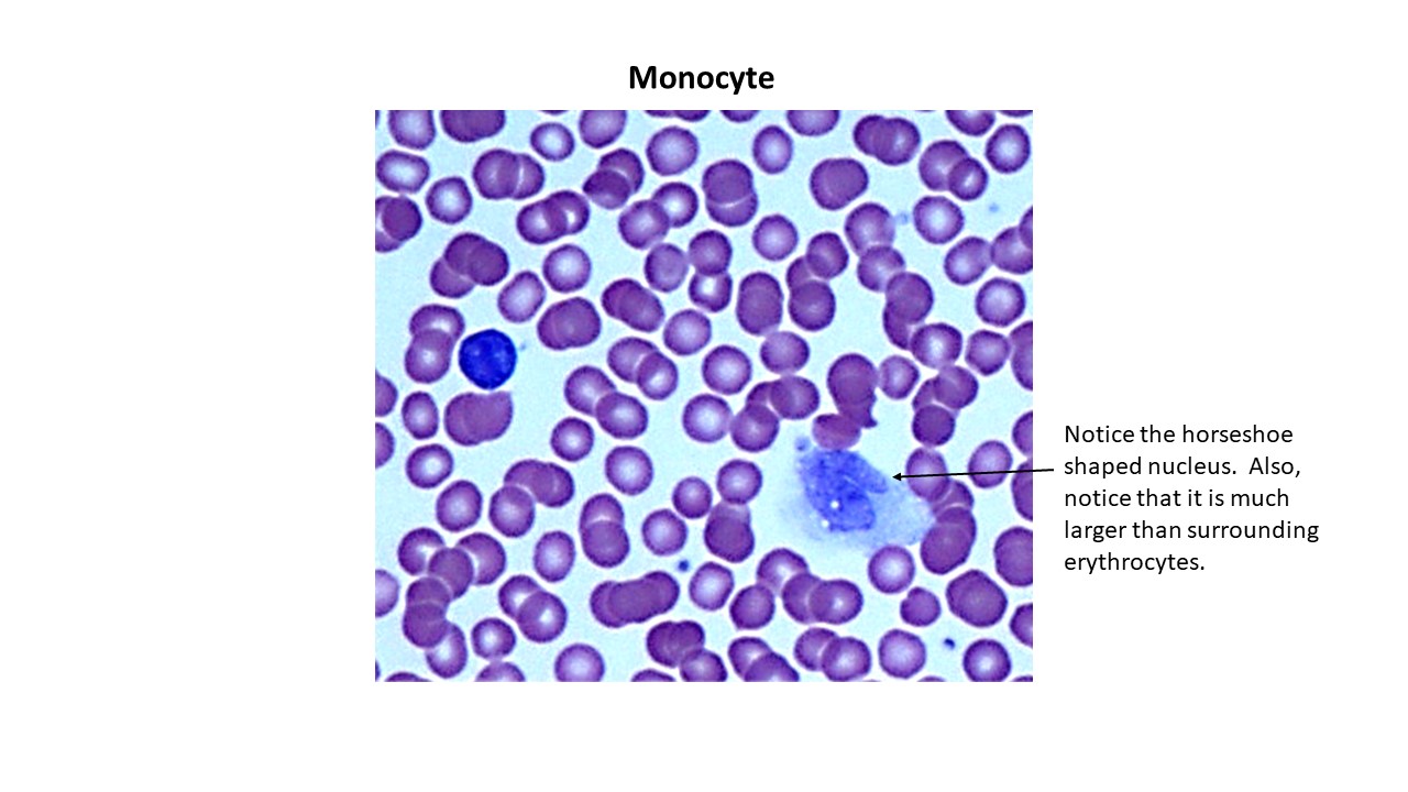
- Monocyte-Monocyte
The Cardiovascular System: Heart
Heart Models
Right Atrium
- Openings of the Superior and Inferior Vena Cava
- Opening of the Coronary Sinus
- Right Auricle with Pectinate Muscles
- Fossa Ovalis (also called the oval fossa)
Right Ventricle
- Chordae Tendineae- attached to the cusps of the tricuspid valve
- Papillary Muscles- chordae tendineae are anchored to these muscles
- Trabeculae Carneae- irregular, muscular ridges within the surface of the internal wall
Left Atrium
- Mainly smooth walled chamber
- Left auricle with Pectinate Muscles- there are fewer muscles compared to the right auricle
Left Ventricle
- Chordae Tendineae- attach to the cusps of the bicuspid valve
- Papillary Muscles- chordae tendineae are anchored to these muscles
- Trabeculae Carneae- irregular, muscular ridges within the surface of the internal wall
- Myocardium is usually 3 times thicker in the left ventricular wall when compared to the right ventricular wall
Heart Valves
- Tricuspid (right atrioventricular valve)-between right atrium and right ventricle
- Bicuspid (aka left atrioventricular valve, mitral valve)-between left atrium and left ventricle
- Aortic semilunar valve- located between the left ventricle and aorta. When this valve opens, blood flows into to the ascending aorta.
- Pulmonary semilunar valve- opens to the pulmonary trunk artery
Coronary Arteries
- Right Coronary Artery (RCA)
- Right Marginal artery (also known as Marginal branch of RCA)
- Posterior interventricular artery (PIA) lies within the posterior interventricular sulcus
- Left Coronary Artery (LCA)
- Circumflex artery
- Anterior interventricular artery (AIA) lies within the anterior interventricular sulcus
Cardiac Veins
- Great Cardiac vein- Accompanies AIA
- Middle Cardiac vein- Accompanies PIA
- Small Cardiac vein- Accompanies Right Marginal artery of RCA (Marginal branch of RCA)
Sulci
- Anterior interventricular sulcus
- Posterior interventricular sulcus
- Coronary sulcus
Coronary Sinus
- This vessel can be seen on the posterior aspect of the heart within the coronary sulcus. It drains low oxygenated blood from the cardiac veins into the right atrium
Large Blood Vessels
- Superior Vena Cava
- Inferior Vena Cava
- Aorta- branches of the aorta are the ascending, aortic arch, thoracic and abdominal
- Pulmonary Trunk- divides into right and left pulmonary arteries. Delivers deoxygenated blood to lungs.
- Pulmonary Veins (2 right Pulmonary and 2 left pulmonary veins). Note that the color of these veins will be red on the models because they carry oxygenated blood from the lungs to the left atrium of the heart.
Pig Heart–View the Dissection Videos of the heart. Also, view the pictures and labels of the dissected pig heart in the file: Lab Practical 3 Review Pictures. These will be the structures you need to know for the lab practical.
- Sulci
- Anterior interventricular sulcus- contains the Anterior Interventricular artery, Great Cardiac vein
- Posterior interventricular sulcus- contains the Posterior Interventricular artery, Middle Cardiac vein
- Coronary sulcus- contains the Coronary sinus on the posterior side of the heart.
- Coronary Sinus
- Pulmonary Trunk Artery
- Aorta
- Chambers of the Heart
- Right Atrium
- Notice the smooth walled portion
- Pectinate Muscles-the rough walled portion found in the auricle
- Openings of Superior and Inferior Vena Cava
- Look for fossa ovalis. Since it is hard to see, it will not be on the practical
- Left Atrium
- Smooth walled portion
- Pectinate Muscles-the rough walled portion found in the auricle
- Right and Left Ventricles
- Be able to tell the difference between each ventricle. The myocardium of the left ventricular wall is much thicker than the left ventricle.
- Ventricles are all rough walled-called the trabeculae carneae
- Papillary muscles
- Chordae tendineae-attached to the valve cusps of bicuspid and tricuspid valves.
- Right Atrium
- Valves of the Pig Heart
- Tricuspid (right atrioventricular valve)-between right atrium and right ventricle
- Bicuspid (left atrioventricular valve, mitral valve)-between left atrium and left ventricle
- Aortic semilunar valve- located between the left ventricle and aorta. Opens to the ascending aorta
- Pulmonary semilunar valve- opens to the pulmonary trunk artery
The Cardiovascular System: Vessels and Circulation
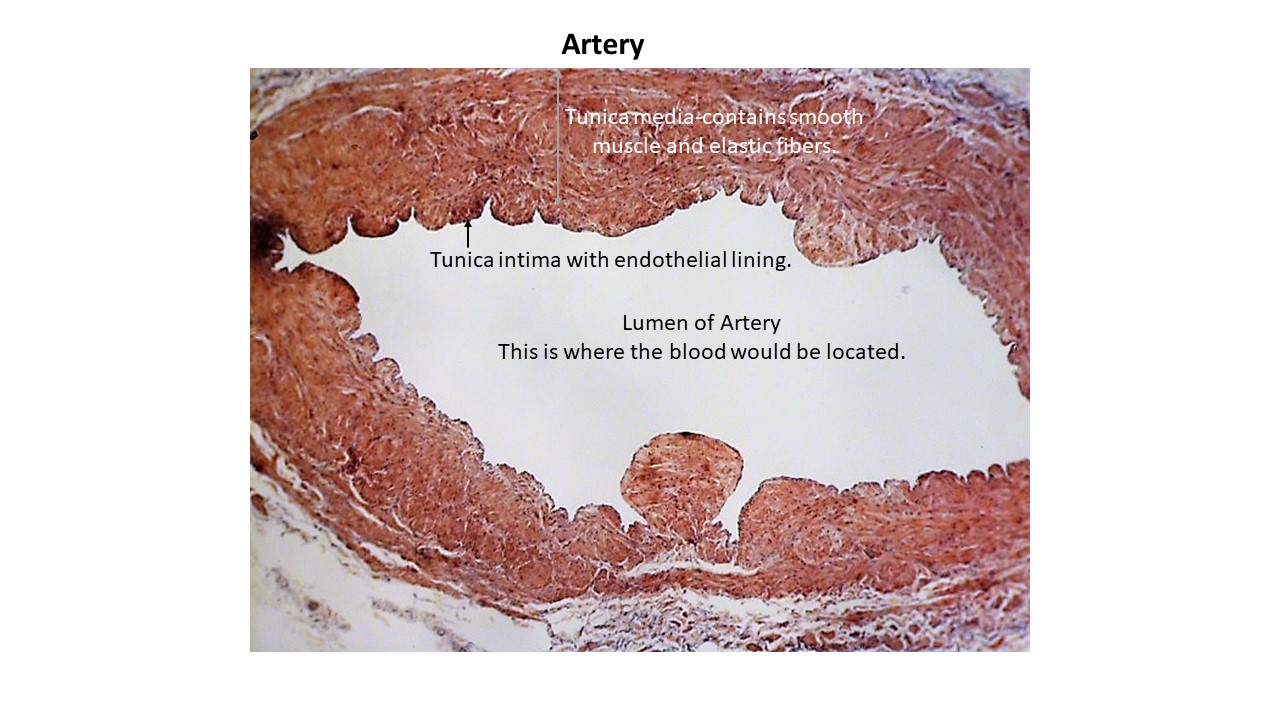
Arteries- blood vessels that transport blood away from the heart
Aorta- Branches
- Ascending Aorta
- Right and left coronary arteries
- Aortic Arch
- Brachiocephalic trunk that branches into right common carotid artery and right subclavian artery.
- Left common carotid artery
- Left subclavian artery
- Thoracic Aorta
- Intercostal
- Abdominal Aorta
- Renal arteries (paired)
- Celiac Trunk artery (unpaired)
- Superior Mesenteric artery (unpaired)
- Inferior Mesenteric artery (unpaired)
Subclavian Branches
- Becomes Axillary artery in axillary region
- Becomes Brachial artery in the brachial region
- Divides into the Radial artery and Ulnar artery
Common Carotid Branches
- External Carotid artery
- Internal Carotid artery
Abdominal Aorta Branches
- Abdominal Aorta divides into the Left Common Iliac and Right Common Iliac arteries
- External Iliac artery
- Internal Iliac artery
- Femoral artery
- Popliteal artery
- Anterior Tibial artery- continues to become the Dorsalis Pedis artery to supply the dorsum of the foot. The Dorsalis Pedis artery is usually not visible on models and will not be on the lab practical.
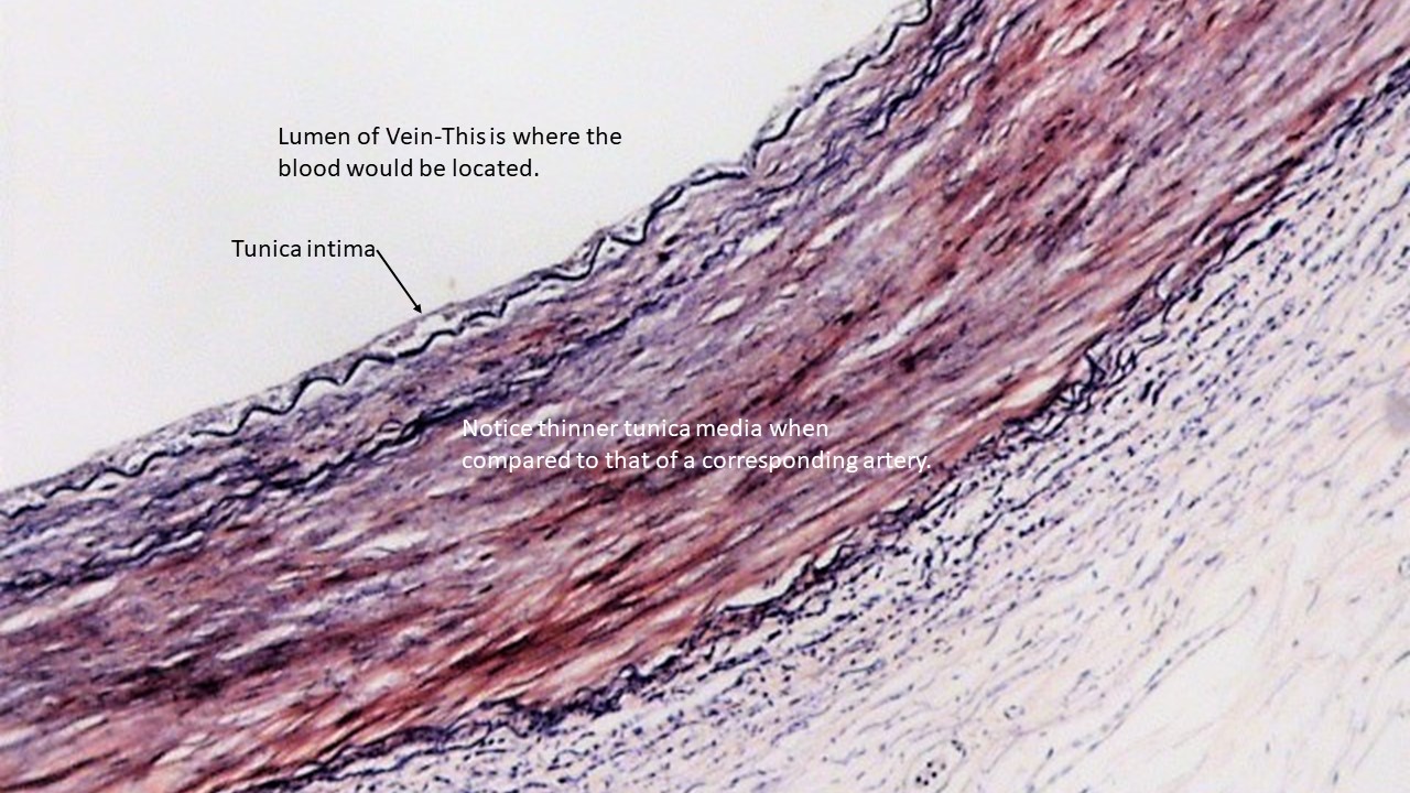
Veins- blood vessels that transport blood to the heart
Superior Vena Cava
- Right and Left Brachiocephalic veins join to become the Superior Vena Cava
Inferior Vena Cava
- Right and Left Common Iliac veins join to form the Inferior Vena Cava
Coronary Sinus
- Receives blood from the Great, Middle and Small Cardiac veins. Drains blood into the right atrium of the heart. The coronary sinus is visible on the view of the posterior heart.
Veins of the Head and Neck
- External Jugular vein (lays on top of the Sternocleidomastoid muscle)
- Internal Jugular vein
Veins of the Upper Limb
- Brachiocephalic vein- the left and right brachiocephalic veins joins to form the Superior Vena Cava
- Subclavian vein- joins with the jugular vein to form the brachiocephalic vein
- Basilic vein
- Cephalic vein (runs in a grove associated with the deltoid muscle)
- Brachial vein
- Median Cubital vein- connects the basilic and cephalic veins
- Radial vein
- Ulnar vein
Veins of the Lower Limb
- Common Iliac vein- the right and left common iliac veins join to form the inferior vena cava
- External Iliac vein- the left and right external iliac veins join to form the common iliac veins
- Internal Iliac vein- joins with the external iliac vein to form the common iliac vein
- Femoral vein- drains into the external iliac vein
- Great Saphenous vein- drains into the femoral vein