
5 Lab Practical 5
Introduction and Learning Objectives
Lab Practical 5 contains structures from the following chapters in your textbook
- Chapter 20-The Endocrine System
- Chapter 27-The Urinary System
- Chapter 28-The Reproductive System
Introduction
In this section, we will first study the endocrine system. What makes this system so distinctive is that organs of the endocrine system are not connected physically, in many situations like other organ systems, but are connected, nonetheless. How, you might ask? The organs of the endocrine system are connected by our cardiovascular system and, more specifically, our blood, which we covered in Lab Practical 3. Endocrine organs release chemicals known as hormones, and hormones can travel throughout our body and reach what are known as target organs by traveling in our bloodstream. Pretty neat, right?
After studying the endocrine system, we will cover the female and male reproductive systems. What makes these two systems unique is that they are both critical for reproduction and produce the cells that are responsible for reproduction, i.e. the oocyte and the sperm. Both also contain organs that are considered endocrine organs because of the production of hormones; the ovaries produce female hormones and the testes produce male hormones. In addition, much of the male’s reproductive ductwork is also used by the male’s urinary tract and the reason that we also study the urinary system in this section.
So, although the endocrine system, reproductive system and urinary system may not seem all that connected on the surface, they are very much interconnected and the rationale behind covering these systems together. As always, we hope that we have stimulated your interest and that you are ready to learn about these miraculous organ systems!
Course Learning Outcomes (CLO)
Identify and Understand Most Major Body Systems
Assessments used:
- Quizzes in the form of self-check assessments (formative)
- Lab Practical Exam (summative)
Module Learning Outcomes
Endocrine System:
- Identify the structural components of the endocrine system (1) (2)
- Describe the function(s) of each endocrine organ and the hormone(s) produced by each within the endocrine system (1)
Urinary System:
- Identify the organs of the urinary system and the function(s) of each of the major organs in this system (1) (2)
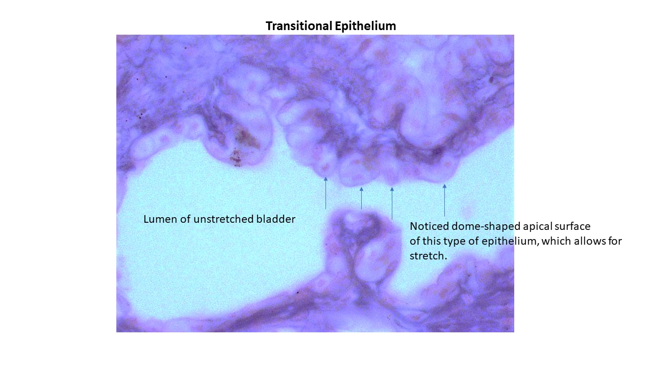
- Identify transitional epithelium and the detrusor muscle tissue slides under the microscope (2)
- Describe the flow of urine from filtration in the glomerular capsule to the urethra (1)
Reproductive System:
- Identify the structural components of the female and male reproductive systems and list their functions (1) (2)
- Identify the male accessory reproductive organs and describe the functions of each (1) (2)
Additional Knowledge:
- Identify the key microscopic features of the endocrine tissue slides under the light microscope (2)
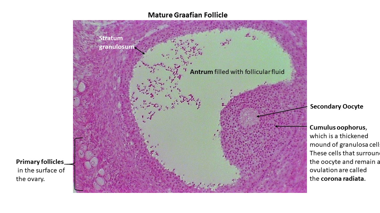
- Identify the key microscopic features of the Graafian ovarian follicle under the light microscope (2)
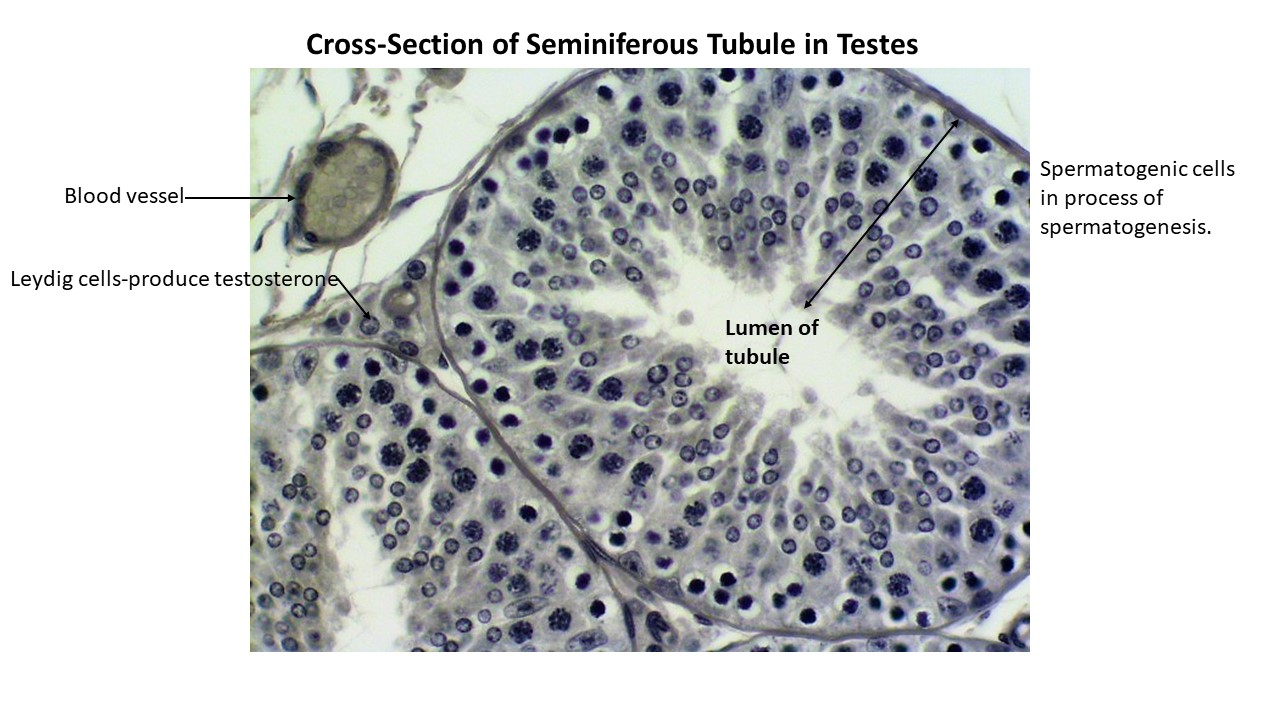
- Identify the key microscopic features of teh seminiferous tubules under the light microscope (2)
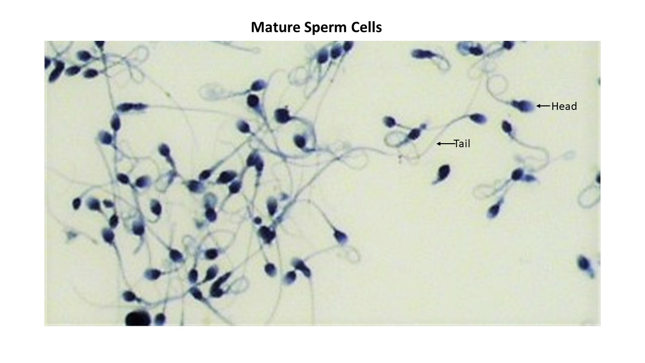
- Identify the key microscopic features of the human sperm under the light microscope (2)
First: Watch Dr. Gannon’s Videos and a Dissection Videos
- The videos will introduce you to the models and, associated structures, that you are expected to learn for Lab Practical 5.
- You are encouraged to watch the videos multiple times as part of your preparation for Lab Practical 5.
Playlists
Endocrine System Playlist
Urinary System Playlist
Reproduction System Playlist
Second: Lab Practical 5 Review Slides
Review the documents linked below. Be able to identify every structure that is labelled in the documents.
- Hint: Learn the material by hand drawing each model, illustration and histology slide and adding the labels for each on your drawing.
Third: Quick Check Assessments (Formative Assessment)
- First, watch the videos linked at the top of each assessment.
- Then, take the assessments in the form of a quiz. The quizzes can be taken multiple times to help you prepare for the lab practical.
Urinary System Anatomy of the Kidney
Female Reproductive System Models
Male Reproductive System Models
Structures To Know Lab Practical 5
- Rewrite the names of every structure on this list to help with retention, recall and spelling.
- Note: Structures may be found on multiple models, illustrations and histology slides.
Endocrine System
- Brain
- Hypothalamus
- Pituitary gland
- Pineal gland (posterior epithalamus)
- Thyroid gland- on anterior surface of trachea (windpipe)
- Parathyroid glands- on posterior surface of thyroid gland (usually 4 small glands)
- Adrenal glands- located on top of each kidney
- Adrenal cortex- outer layer
- Adrenal medulla- inner layer
- Pancreas
Histology Slides– Be able to identify the following tissues of endocrine glands.
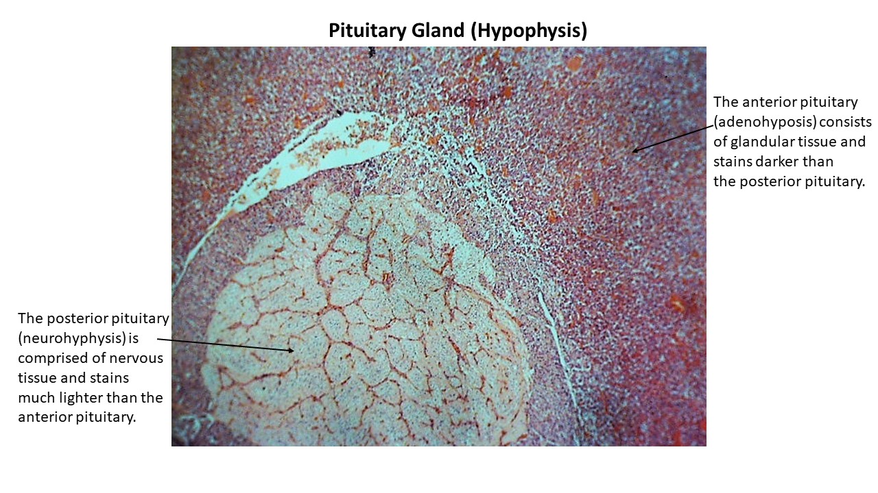
- Pituitary gland: anterior pituitary, posterior pituitary
- Pituitary Gland
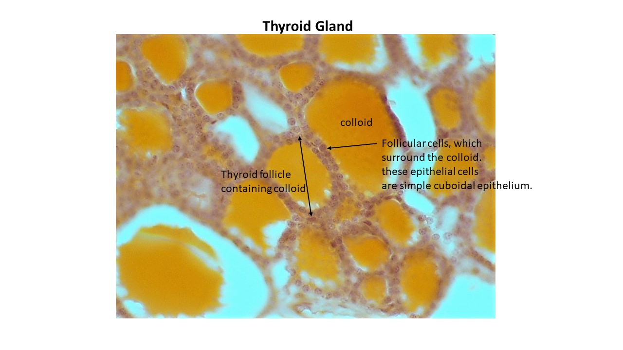
- Thyroid gland
- Thyroid follicle
- Colloid within follicle
- Follicular cells
- Thyroid Gland
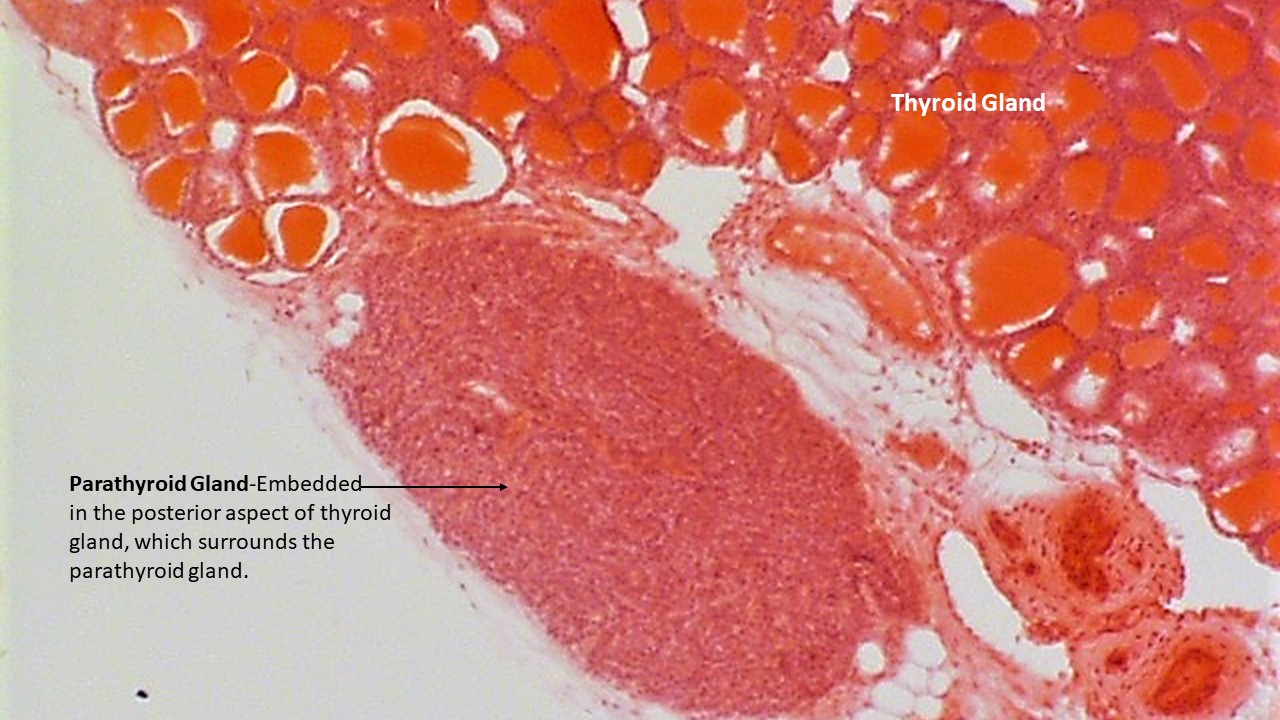
- Parathyroid glands- usually viewed in a tissue section including a section of the thyroid
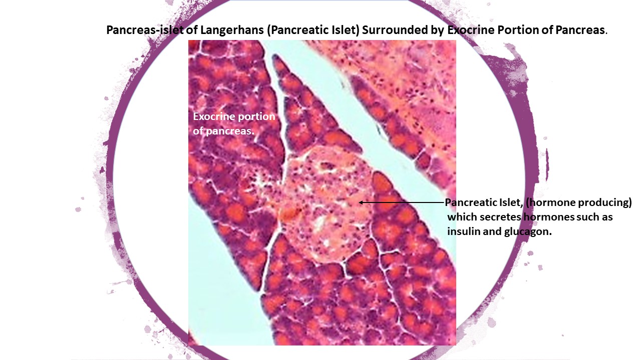
- Pancreas
- Pancreatic Islet
- Pancreas with Islet of Langerhans (Pancreatic Islet)
Urinary System
Kidney Models and Nephron Models
- Renal Cortex- outer layer of the kidney
- Renal Medulla- inner layer of the kidney
- Renal Pyramids
- Renal Columns
- Renal Papilla- at the end of the renal pyramid
- Nephrons
- Minor Calyces (Calyx – singular form)
- Major Calyces
- Renal Pelvis
- Ureters
- Renal Artery
- Afferent Arterioles
- Efferent Arterioles
- Renal Vein
- Renal Corpuscle
- Renal Tubule
- Glomerulus
- Glomerular Capsule (also known as Bowman’s Capsule)
- Proximal convoluted tubule
- Ascending Limb of the Nephron Loop
- Descending Limb of the Nephron Loop
- Distal Convoluted Tubule
- Collecting Ducts
- Urinary Bladder
- Urinary Bladder- Internal structure
- Ureteral openings and Internal Urethral Opening à form trigone of bladder
- Detrusor muscle
Pig Kidneys
- Renal Cortex
- Renal Medulla
- Renal Pyramids
- Renal Columns
- Minor Calyces (Minor calyces are just beneath the renal pyramids)
- Major Calyces (seen just below the junction of 2-3 minor calyces which empty into a major calyx)
- Hilum- area of the kidney
- Renal Pelvis- seen just before the ureter
- Ureters- light colored tissue; note that the ureters are not visible in the dissection pictures
- Renal Artery- not visible in the dissection pictures
- Renal Vein- not visible in the dissection pictures
Histology Slides- Be able to identify the following tissues of the urinary system.
- Bladder-Be able to identify the bladder lined with transitional epithelium.
- Bladder with transitional epithelium.
The Reproductive System
Male Reproductive System
- Scrotum
- Testes
- Epididymis
- Spermatic cord
- Ductus deferens
- Ampulla of ductus deferens
- Seminal vesicles
- Ejaculatory duct
- Male urethra- 3 parts
- Prostatic urethra
- Membranous urethra
- Penile or Spongy urethra
- Prostate gland
- Bulbourethral glands
- Penis
- Body
- Glans penis
- Corpora cavernosa (corpus cavernosum- singular form)
- Corpus spongiosum
Female Reproductive System
- Ovaries
- Ovarian ligament
- Mesovarium (Mesosalpinx)
- Broad ligament
- Suspensory ligament
- Uterine tubes or oviduct (also known as Fallopian tubes)
- Fimbriae
- Infundibulum
- Ampulla
- Isthmus
- Uterus
- Fundus
- Body
- Cervix
- Vagina
- Mammary glands- view on the instructional video of the female reproductive system
- Areola
Histology Slides- Be able to identify the structures on the ovary and testes slide. Also, be able to identify mature sperm cells and the structures labelled on this slide.
- Ovary– Mature Graafian follicle.
- Mature Graafian follicle
- Testes-Seminiferous Tubule
- Seminiferous Tubule
- Mature Sperm Cells