
4 Lab Practical 4
Introduction and Learning Objectives
Lab Practical 4 contains structures from the following chapters in your textbook
- Chapter 14-NervousTissue
- Chapter 15-Brain and Cranial Nerves
- Chapter 16-Spinal Cord and Spinal Nerves
Introduction
Our nervous system is an amazing and, somewhat, mysterious system. It has been described as the last unknown frontier of the human body because some functions of the nervous system remain elusive. However, considerable knowledge has been gained over recent decades and scientists have a much better understanding of how the nervous system functions to maintain internal homeostatic conditions and serve as a major communication system for our body.
Let us focus on the communication function, for a moment, and consider a real-world example of how the nervous system serves to control communication by processing external stimuli and, in response, make changes inside of our body.
Have you ever walked into a dimly lit room? Without thinking about it consider what happened internally in response to the low light levels in the room. Your pupils likely dilated to allow additional light to get to the photoreceptors of your eye so that you could see better. In this example, your nervous system responded to an external stimulus (change in light) and was able to respond internally (pupillary dilation) because of the communication processes that are part of your nervous system.
In this section we will learn how nervous tissue, such as neurons and neuroglial cells, the brain, cranial nerves, spinal cord, and spinal nerves all work together for the purpose of communication and maintenance of homeostatic conditions. Our nervous system is remarkable in its ability to eloquently handle and respond to the barrage of stimuli that it receives. We hope that you will agree after completing your study of the nervous system.
Course Learning Outcomes (CLO)
Identify and Understand Most Major Body Systems
Assessments used:
- Quizzes in the form of self-check assessments (formative)
- Lab Practical Exam (summative)
Module Learning Outcomes
Successful completion of this module demonstrates that you have met the following Module Level Outcomes (MLO). The numbers at the end of each MLO corresponds to the assessment used to measure attainment of the Course Learning Outcome (CLO).
Brain Models:
- Identify the different parts of the brain and their functions (1) (2)
Spinal Cord Models:
- Identify the different parts of the spinal cord and their functions (1) (2)
- Describe the structure and function(s) of the spinal cord (1)
- Describe the arrangement of white and gray matter within the spinal cord (1)
- Identify the three spinal meninges and the spaces associated with the meninges (1).
- Identify the spinal nerve plexuses and the functions in each plexus (1)
Brain Stem Model:
- Identify the twelve cranial nerves and their functions (1) (2)
Additional Knowledge:
- Identify the key microscopic features of nervous tissue and the spinal cord cross section using a light microscope. (2)
First: Watch Dr. Gannon’s Videos and Dr. Zimmerman’s Dissection Videos
- The videos will introduce you to the models and, associated structures, that you are expected to learn for Lab Practical 4.
- You are encouraged to watch the videos multiple times as part of your preparation for Lab Practical 4.
Playlists
Nervous System Playlist
Dr. Zimmerman’s Dissection Videos
Sheep Brain Dissection-Exterior Structures
Sheep Brain Dissection-Interior Structures
Second: Lab Practical 4 Review Slides
Review the documents linked below. Be able to identify every structure that is labelled in the documents.
- Hint: Learn the material by hand drawing each model, illustration and histology slide and adding the labels for each on your drawing.
Third: Quick Check Assessments (Formative Assessment)
- First, watch the videos linked at the top of each assessment.
- Then, take the assessments in the form of a quiz. The quizzes can be taken multiple times to help you prepare for the lab practical.
A215 Spinal Cord-Reflex Arc Model
A215 Spinal Cord Cervical Region
A215 Brain Stem and Cranial Nerves
Structures To Know Lab Practical 4
- Rewrite the names of every structure on this list to help with retention, recall and spelling.
- Note: Structures may be found on multiple models, illustrations and histology slides.
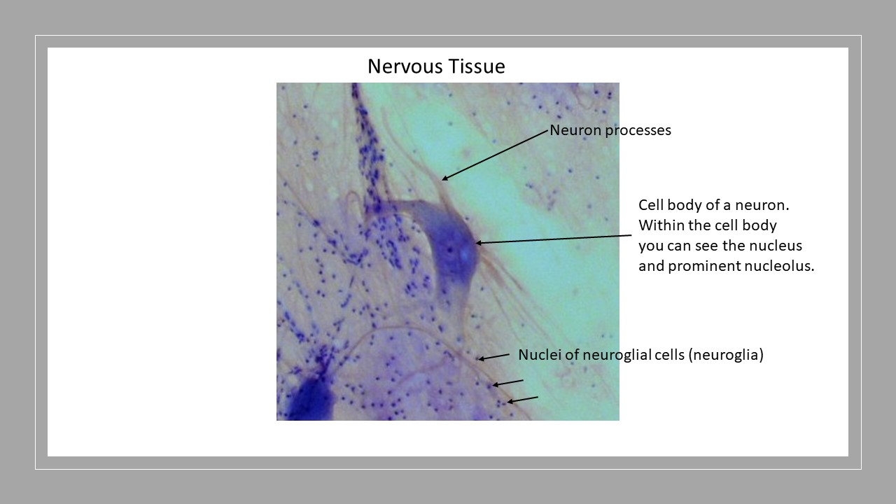
Nervous Tissue
Neuron: Review the slide of the nervous tissue and the labelled neuron.
- Nervous Tissue
- Neuron processes
- Cell body (soma)
- Nucleus
- Nucleolus
The Spinal Cord and Spinal Nerves
Plexuses and Nerves- Identify these structures on the nervous system flat model and the large spinal cord model
- Cervical plexus
- Phrenic nerve
- Brachial plexus- you do not have to know the lateral, posterior and medial cords for the virtual lab exam; however, be sure to know the location of the following nerves:
- Axillary
- Musculocutaneous
- Radial
- Ulnar
- Median
- Thoracic Region
- Intercostal Nerves
- Lumbar plexus
- Iliohypogastric nerve
- Ilioinguinal nerve
- Genitofemoral nerve
- Femoral nerve
- Lateral Femoral Cutaneous nerve
- Obturator nerve
- Sacral plexus
- Sciatic Nerve
Other Structures on the Large Spinal Cord:
- Filum Terminale
- Conus Medullaris
- Cauda Equina
Models of the Spinal Cord
- Cross Section of the Spinal Cord Model- Cervical Region. Know all the structures on the key. Learn these structures as there will be 8 questions over this model. These are covered in Dr. Gannon’s videos.
Histology Slide of Spinal Cord: Review the slide of the spinal cord and know the labelled structures.
Cervical and Reflex Arc
- Anterior median fissure
- Posterior median sulcus
- Posterior gray horns
- Anterior gray horns
- Lateral gray horns
- Gray commissure
- Gray matter
- White matter
- White posterior column
- White anterior column
- Dorsal (posterior) root
- Ventral (anterior) root
- Epidural space
- Spinal nerve at the intervertebral foramen
- Dura mater
- Subarachnoid space
- Arachnoid mater
- Subdural space
- Pia mater
- Denticulate ligaments
The Brain and Cranial Nerves
Brain Models: External Structures
Cerebrum- Lobes
- Frontal
- Precentral gyrus; Motor Cortex
- Parietal
- Postcentral gyrus; Somatosensory Cortex
- Temporal
- Occipital
Fissures and Sulci
- Longitudinal fissure
- Transverse fissure
- Central sulcus
- Lateral sulcus
Brain Models: Internal Structures
- Corpus callosum
- Hypothalamus- identify the area of the hypothalamus on the sagittal section of the brain models
- Pituitary gland
- Pineal gland
- Optic chiasm
- Optic nerve
- Thalamus- be able to identify the area of the thalamus
- Mammillary body
- Lateral ventricle
- 4th ventricle
- Subarachnoid space
- Cerebral aqueduct or aqueduct of the midbrain
- Cerebellum: arbor vitae (the white matter), folia or cerebellar cortex (gray matter)
- Midbrain
- Pons
- Medulla oblongata
Dissected Sheep Brain: Watch the sheep brain dissection videos.
External Sheep brain:
- Gyri
- Sulci
- Dura mater
- Pia mater
- Cerebrum: Frontal, Parietal, Temporal, and Occipital lobes
- Olfactory bulbs
- Optic nerve
- Optic chiasm
Dorsal Sheep Brain
- Corpora quadrigemina: superior and inferior colliculi
Sheep Brain Stem:
- Midbrain
- Pons
- Medulla oblongata
Sheep Brain Cerebellum
- Arbor vitae
- Folia or Cerebellar cortex
Sheep Spinal cord: Central canal
Internal sheep brain
- Corpus callosum
- Choroid plexus in the lateral and 4th ventricles
- Lateral ventricle and the 4th ventricle
- Cerebral aqueduct or aqueduct of the midbrain
- Hypothalamus
- Optic chiasm
- Optic nerve
- Mammillary body
- Pineal gland in the epithalamus
- Cerebellum: arbor vitae (the white matter), folia or cerebellar cortex (gray matter)
- Pons
- Medulla oblongata
Cranial Nerves on the Brain Stem Model
Identify the 12 Cranial Nerves. Know their names and the roman numerals for each. Also, be able to identify all structures on the key.