
6 Lab Practical 6
Introduction and Learning Objectives
Lab Practical 6 contains structures from the following chapters in your textbook
- Chapter 24-The Respiratory System
- Chapter 25-The Digestive System
Introduction
Lab Practical 6 concludes our study of anatomy. In this section we will cover our final chapters, the respiratory and digestive systems.
In concert with the cardiovascular system, our respiratory system is responsible for delivering oxygen to the cells of our body. There are many functions that this system performs, but we will focus on the parts of the respiratory system that serve as passageways for the movement of air into the sacs of our lungs. Like other systems, diseases that impact our respiratory system can significantly and negatively impact our daily lives. Everything from chronic obstructive pulmonary disease to the more recent novel coronavirus known as COVID-19, affect the respiratory system.
The digestive system is a tube with openings on both ends at the oral cavity and anal canal. In utero, the digestive system begins as a tube that twists and dilates to form the oral cavity and esophagus positioned cranially to the rectum and anal canal positioned caudally plus everything inbetween. In this section we will learn the various structures of the digestive system and take a closer look, histologically, at some of the cellular features that allow this system to absorb nutrients across the epithelial cell walls of this system.
We hope that you have enjoyed your study of anatomy and have gained a greater appreciation of the complexity of the human body!
Course Learning Outcomes (CLO)
Identify and Understand Most Major Body Systems
Assessments used:
- Quizzes in the form of self-check assessments (formative)
- Lab Practical Exam (summative)
Module Learning Outcomes
Respiratory Models:
- Identify and locate the organs of the respiratory system. (1) (2)
Digestive Models:
- Identify and locate the organs of the digestive system. (1) (2)
- Identify the accessory organs of the digestive system (2)
- Compare and contrast the four layers of the digestive tract and the characteristics of each layer (1) (2)
Additional Knowledge:
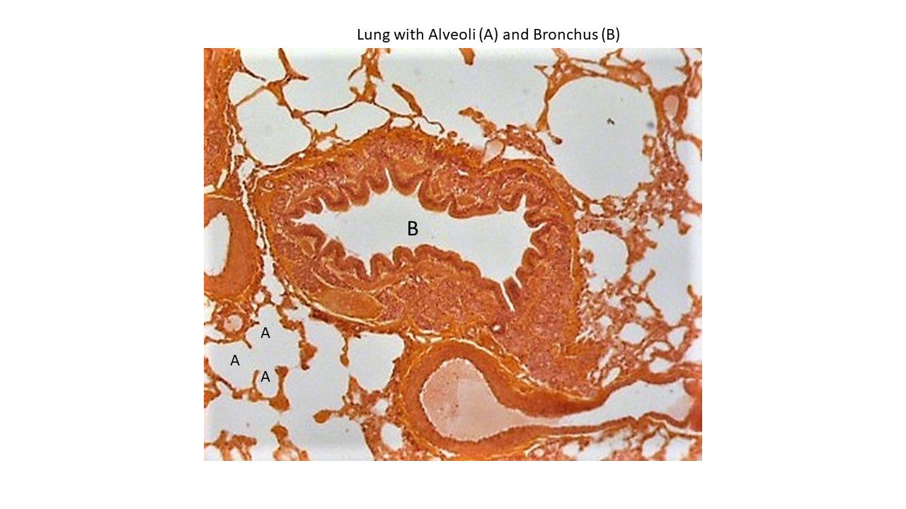
- Identify the key microscopic features of lung tissue with alveoli under the light microscope (2)
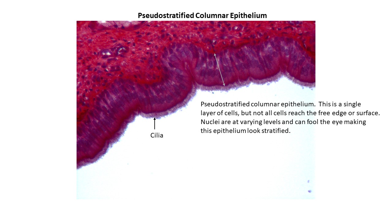
- Identify the key microscope features of the trachea with pseudostratified columnar epithelium and cilia under the light microscope (2)
First: Watch Dr. Gannon’s Videos and a Dissection Videos
- The videos will introduce you to the models and, associated structures, that you are expected to learn for Lab Practical 6.
- You are encouraged to watch the videos multiple times as part of your preparation for Lab Practical 6.
Playlists
Respiratory Playlist
Digestive Playlist
Second: Lab Practical 6 Review Slides
Review the documents linked below. Be able to identify every structure that is labelled in the documents.
- Hint: Learn the material by hand drawing each model, illustration and histology slide and adding the labels for each on your drawing.
Third: Quick Check Assessments (Formative Assessment)
- First, watch the videos linked at the top of each assessment.
- Then, take the assessments in the form of a quiz. The quizzes can be taken multiple times to help you prepare for the lab practical.
A215 Digestive System Overview
A215 Digestive System Small Intestines
A215 Respiratory System Part 1
A215 Respiratory System Part 2
Structures To Know Lab Practical 6
- Rewrite the names of every structure on this list to help with retention, recall and spelling.
- Note: Structures may be found on multiple models, illustrations and histology slides.
Lab Practical 6
Respiratory System
Respiratory Models
- Paranasal Sinuses
- o Frontal
- o Sphenoidal
- Nasal Cavity and Nose
- o External Nares
- o Internal Nares
- o Superior, Middle and Inferior Nasal Conchae
- Pharynx- commonly called the throat. Originates posterior to the nasal and oral cavity and extends to the level of the beginning of the larynx and esophagus
- o Nasopharynx- lined with pseudostratified ciliated columnar epithelium.
- Location of pharyngeal tonsils
- Opening of auditory (Eustachian) tubes or pharyngotympanic tubes
- o Oropharynx- lined with nonkeratinized stratified squamous epithelium.
- Palatine tonsils- located on the posterior, lateral wall of the oral (buccal) cavity
- Lingual tonsils- located at the base of the tongue
- o Laryngopharynx- lined with nonkeratinized stratified squamous epithelium.
- Continues inferiorly to the start of the esophagus located posteriorly and larynx located anteriorly
- Uvula- extension of the soft palate which functions to close off the nasopharynx when swallowing. Prevents food and liquids from entering the nasal cavity
- Larynx
- o Thyroid cartilage
- o Cricoid cartilage
- o Epiglottis
- o Glottis
- o Arytenoid cartilage
- o True vocal cords (vocal folds)
- o False vocal cords (vestibular folds)
- Trachea
- o Tracheal (C-shaped) rings- these rings are open in the back to permit distension of the esophagus which lies posterior to the trachea.
- o Trachealis muscle- binds to the C-shaped rings of the trachea
- Bronchi- project inferiorly and laterally toward each lung.
- o Right Primary Bronchus- wider and shorter than the left primary bronchus
- o Left Primary Bronchus- narrow and longer than the right primary bronchus
- Lungs
- o Nasopharynx- lined with pseudostratified ciliated columnar epithelium.
-
- Both Lungs-apex (superior portion of lungs), base (inferior portion of lungs) and hilum (bronchi and pulmonary vessels enter here)
- Right Lung-3 lobes: superior, middle and inferior lobes with horizontal and oblique fissures
- Left Lung-2 lobes: superior and inferior lobe with oblique fissure
- Left Lung-Cardiac Notch- an indented region
Histology Slides
- Trachea-Trachea with Pseudostratified Columnar Epithelium and Cilia
- Lung Tissue with Alveoli-Lung Tissue
Digestive System
Digestive System Models
- Oral or Buccal Cavity
- Lips
- Hard palate- formed by the palatine process of the maxilla and part of the palatine bone
- Soft palate- posteriorly located. Uvula extends inferiorly from the soft palate. When swallowing the soft palate and uvula raise to close off the nasopharynx. Prevents food and liquids from entering the nasal region
- Salivary Glands
- Parotid glands
- Submandibular glands
- Sublingual glands
- Tongue
- Esophagus
- Stomach
- o Greater curvature, lesser curvature
- o Cardia
- o Fundus
- o Body
- o Pylorus
- o Pyloric Sphincter
- o Rugae- folds in the mucosa
- Pancreas
-
- Pancreatic duct
- Accessory pancreatic duct
- Hepatopancreatic ampulla (sphincter)
- Head, body and tail of pancreas
- Liver
-
- Right and left lobes
- Gallbladder-located under the right lobe-cystic duct empties gallbladder
- Right and left hepatic ducts and common hepatic duct
- Common bile duct
- Mesenteries- folds of peritoneum
- Greater Omentum- extends inferiorly from the greater curvature of the stomach over the abdominal region. Be able to identify this on several of the models
- Mesocolon- fold of peritoneum which attaches to the large intestine
- Small Intestine
-
- Duodenum- the first part of the small intestine
- Jejunum and Ileum- you won’t be able to tell where jejunum ends and ileum begins; however know that the jejunum is the upper part of SI and the ileum is the lower part of SI.
- Ileocecal Valve- sphincter muscle allowing flow of materials into the large intestine
- Small Intestine Villi Model- see the instructional video of this model and the labeled model in the review powerpoint. Know all labeled parts.
- Large Intestine
-
- Cecum
- Appendix
- Ascending, Transverse, Descending Colon, Sigmoid Colon
- Rectum
- Haustra
- Taeniae coli