
2 Lab Practical 2
Introduction and Learning Objectives
Lab Practical 2 includes structures covered in the following chapters of your textbook:
-
- Chapter 6-Cartilage and Bone
- Chapter 7-Axial Skeleton
- Chapter 8-Appendicular Skeleton
- Chapter 10-Muscle Tissue Organization
- Chapter 11-Axial Muscles
- Chapter 12-Appendicular Muscles
Introduction
We are now beginning our journey of the remaining organ systems. In this section, we will cover the skeletal system and the muscular system. Collectively, these two systems are often referred to as the musculoskeletal system.
Consider all the major functions of our skeletal system including protection of our internal organs, production of blood cells by red bone marrow, and support of our body. Without your skeletal system, you would be unable to stand, sit or move. Anyone that has broken a bone understands the limitations that can present following a bone fracture.
Similarly, can you imagine performing everyday tasks without muscle tissue? In this section we will learn about the three types of muscle tissue: skeletal, smooth, and cardiac muscle. Skeletal muscle allows us to do everything from sit, stand, write, walk, and talk. Smooth muscle is critical for maintaining many functions that we do not consciously think about such as maintaining blood pressure, movement of foods and liquids through our digestive tract and even creating goose bumps! And, of course, cardiac muscle is essential to the contraction of our heart and pumping of blood throughout out body.
In this section, we will explore our musculoskeletal system, learn more about the microscopic features of these two systems, and become familiar with some of the key structures on our bones. We hope that we have piqued your interest, so let’s get started!
Course Learning Outcomes (CLO)
Identify and Understand Most Major Body Systems
Assessments used:
- Quizzes in the form of self-check assessments (formative)
- Lab Practical Exam (summative)
Module Learning Outcomes
Successful completion of this module demonstrates that you have met the following Module Level Outcomes (MLO). The numbers at the end of each MLO corresponds to the assessment used to measure attainment of the Course Learning Outcome (CLO).
Bone Model:
- Compare spongy and compact bone (2)
- Identify key structures of bone tissue (1) (2)
Bones
- Identify key regions of a long bone (1)
- Identify the landmarks and the bones of the axial skeleton (1) (2)
- Identify the landmarks and the bones of the appendicular skeleton (1) (2)
Skeletal, Cardiac and Smooth Muscle Models:
- Identify the key microscopic structures of skeletal, cardiac and smooth muscle (2)
Muscle Models:
- Identify the axial skeletal muscles of the body (1) (2)
- Identify the appendicular skeletal muscles of the body (1) (2)
Additional Knowledge
- Identify the key microscopic features of skeletal, cardiac and smooth muscle using a light microscope. (2)
First: Watch Dr. Gannon’s Videos
- The videos will introduce you to the models and, associated structures, that you are expected to learn for Lab Practical 2.
- You are encouraged to watch the videos multiple times as part of your preparation for Lab Practical 2.
Playlists
Bone Tissue
Axial Skeleton
Appendicular Skeleton
Muscular System
Second: Lab Practical 2 Review Slides
Review the documents linked below. Be able to identify every structure that is labelled in the documents.
- Hint: Learn the material by hand drawing each model, illustration and histology slide and adding the labels for each on your drawing.
- Lab Practical 2 Review Pictures-Bones
- Lab Practical 2 Review Pictures-Muscles
Third: Quick Check Assessment Quiz (Formative Assessment)
- First, watch the videos linked at the top of each assessment.
- Then, take the assessments in the form of a quiz. The quizzes can be taken multiple times to help you prepare for the lab practical.
Structures To Know-Lab Practical 2
- Rewrite the names of every structure on this list to help with retention, recall and spelling.
- Note: Structures may be found on multiple models, illustrations and histology slides.
Lab Practical 2 Structure List
The Skeletal System: Cartilage and Bone
- Compact Bone Slide
-
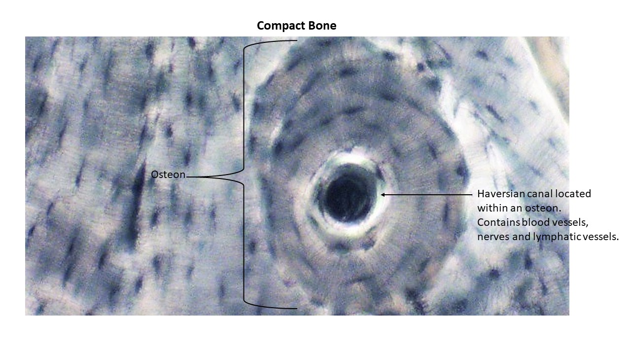
- Compact Bone-Showing Osteon and Haversian Canal
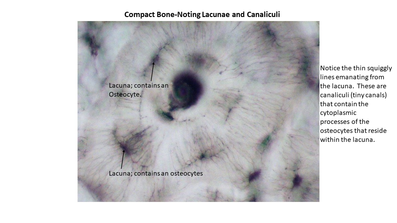
- Compact Bone-Showing Lacunae and Canaliculi
- Haversian (Central canal) of osteon
- Osteon
- Lacunae containing osteocytes within the lacunae
- Canaliculi
- Bone Model
- Osteocytes sitting with lacunae
- Canaliculi
- Lacunae
- Central canal, which contains an artery (red), vein (dark blue), nerve (white), and lymphatic vessel (light blue but not labelled)
- Interstitial lamellae
- Concentric lamellae
- Long Bone
- Epiphysis
- Epiphyseal line-location of the Metaphyseal plate
- Medullary cavity
- Compact bone
- Diaphysis
- Spongy bone in epiphysis
The Skeletal System: Axial Skeleton
- Skull-Sutures
- Coronal suture
- Sagittal suture
- Lambdoid suture
- Squamous suture
- Frontal or Metopic suture (seen on the fetal skull)
- Cranial Bones
o Frontal bone
o Parietal bone (there are 2)
o Occipital bone
o Temporal bone
o Sphenoid
o Ethmoid - Facial Bones
o Maxillae
o Palatine bones
o Nasal bones
o Inferior nasal conchae
o Zygomatic bones
o Lacrimal bones
o Vomer
o Mandible - Occipital Bone-Anatomical Structures-Refer to Figure 6.6 of the textbook
o Foramen magnum-Contains medulla oblongata (inferior portion of brainstem)
o Occipital condyles-Where skull articulates with the first cervical vertebra (C1).
o External occipital protuberance
o Inferior and superior nuchal lines
o Jugular foramen-Several important cranial nerves pass through this foramen as well as the internal jugular vein, which returns blood from the brain to the heart.
o Hypoglossal canals-Hypoglossal nerve (CN XII) passes through this canal and provides motor control to the muscles of the tongue - Parietal Bones-Anatomical Structures-Refer to Figures 6.3 a, b, and Figure 6.6 c of textbook
o Superior and inferior temporal lines-mark the attachment of the temporalis muscle, which is a muscle of mastication (chewing muscle) to aid in closing the mouth. - Frontal Bone-Anatomical Structures-Refer to Figure 6.3b-d of Textbook)
o Supra-orbital margins-mark superior limits of the orbits.
o Supra-orbital foramen or notch-supra-orbital nerve and supra-orbital artery pass through this foramen.
o Frontal sinuses-these vary in size from person to person. Usually begin to develop after age 6, but some of us never develop a frontal sinus. - Temporal Bones-Anatomical Structures-There are many figures to refer to for the temporal bones
o Squamous part-attachment site for muscles of mastication (chewing muscles)
o Zygomatic process of the temporal bone-meets with the temporal process of the zygomatic bone to form the zygomatic arch, also known as the cheekbone.
o Mandibular fossa-articulates with mandible
o Articular tubercle-articulates with mandible
o External acoustic meatus-ear canal
o Petrous part-surrounds and protects the organs of hearing and balance
o Mastoid process-contains mastoid air cells where infections of the respiratory tract can spread leading to mastoiditis.
o Styloid process-attachment site for ligaments and muscles attached to the hyoid bone.
o Stylomastoid foramen-Facial nerve (CNVII) passes through this foramen, which provides innervation to the muscles of facial expression.
o Carotid canal-carotid artery passes through this canal carrying blood from the heart to the brain.
o Internal acoustic meatus-a passageway for blood vessels and nerves to the inner ear. - Sphenoid Bone-Looks like a giant bat or butterfly. Anatomical Structures-Refer to Figures 6.4 and 6.9a of the textbook
o Greater wing
o Lesser wing
o Hypophysial fossa-cradles pituitary gland
o Sella turcica-resembles a “Turkish saddle”
o Optic canal
o Optic groove-allows for passage of the Optic Nerves (CNII)
o Foramen rotundum
o Foramen ovale
o Foramen spinosum
o Pterygoid processes-attachment site for muscles of mastication (chewing muscles) - Ethmoid Bone-Anatomical Structures-There are many figures to refer to for the ethmoid bone
o Cribriform plate, which contains cribriform foramina-These openings allow for passage of the olfactory nerves.
o Crista galli-looks like a rooster’s head dress-falx cerebri attaches here
o Superior nasal conchae
o Middle nasal conchae
o Perpendicular plate-forms upper part of nasal septum
Bones of the Face (Facial Bones) - The Maxillae-Anatomical Structures-There are many figures to refer to for the maxillae
o Alveolar processes-contain upper teeth
o Infra-orbital foramen
o Maxillary sinuses-largest sinuses
o Palatine processes-forms anterior portion of hard palate - Palatine bones-form posterior part of hard palate
- Inferior Nasal Conchae
- Zygomatic Bones
o Temporal process of zygomatic bone-articulates with zygomatic process of temporal bone to form zygomatic arch (cheekbone - Lacrimal bones-smallest facial bone
o Lacrimal groove-leads to nasolacrimal canal - Vomer-forms inferior portion of nasal septum
- Mandible
o Body
o Rami of the mandible
o Angle of the mandible
o Head of the mandible
o Condylar process
o Coronoid process-temporalis muscle inserts onto mandible
o Mental foramina-passage of mental nerve
o Mandibular notch-depression between condylar and coronoid process
o Alveolar part-contains lower teeth
o Mandibular foramen-passage of inferior alveolar nerve - Hyoid bone-Does not articulate with any other bones of the skeleton. Refer to Figure 6.17 in textbook
o Greater and lesser horn - Vertebral Column
o Vertebral Regions-Cervical, Thoracic, Lumbar, Sacral, and Coccygeal-Refer to Figure 6.19 in textbook - Vertebra
o Vertebral body
o Spinous process
o Transverse process
o Pedicle
o Superior and Inferior articular processes
o Vertebral foramen-for passage of spinal cord
o Intervertebral foramen-for passage of spinal nerves
o Intervertebral disc - Sternum
- Manubrium
- Body
- Xiphoid process
- Ribs
- True ribs 1-7
- False ribs 8-12
- Floating ribs-11 and 12
- Costal groove
The Skeletal System: Appendicular Skeleton
Pectoral Girdle
- Clavicle-Noticed that it is “S” shaped.
-
- Acromial end-articulates with the scapula at the acromioclavicular joint
- Sternal end-articulates with the manubrium of the sternum at the sternoclavicular joint
- Scapula
- Acromion
- Glenoid cavity
- Coracoid process
- Suprascapular notch
- Subscapular fossa
- Spine-posterior view
- Supraspinous fossa-posterior view
- Infraspinous fossa-posterior view
Upper Limbs
- Humerus
- Greater tubercle
- Lesser tubercle
-
- Head
- Anatomical neck
- Intertubercular sulcus
- Surgical neck
- Deltoid tuberosity
- Lateral and medial epicondyle
- Capitulum
- Trochlea
- Olecranon fossa
- Radius
- Head of radius-noticed how the head is flat
- Radial styloid process
- Ulna-Looks like an ice cream scoop on proximal side
- Olecranon
- Trochlear notch
- Ulnar styloid process
- Carpal Bones
- Proximal row from lateral to medial-scaphoid, lunate, triquetrum, pisiform
- Distal row from lateral to medial-trapezium, trapezoid, capitate, hamate. Notice that the hamate has a “hook”.
- Metacarpal bones-Notice that they are numbered from I-V with I situated adjacent to the phalanges that make up the thumb.
- Phalanges-Notice that there is a proximal, middle, and distal phalanx except for the thumb, which only has a proximal and distal phalanx.
Pelvic Girdle
- Hip bones, Sacrum and Coccyx
- Acetabulum for articulation with the head of the femur
- Obturator foramen
- Pubic symphysis
- Sacro-iliac joint
- Sacrum
- Sacral foramina
- Sacral promontory
- Ilium
- Iliac crest
- Iliac fossa
- Greater sciatic notch-for passage of sciatic nerve
- Pubis
- Superior pubic ramus
- Inferior pubic ramus
- Ischium
- Spine of ischium
- Lesser sciatic notch
- Ischial tuberosity
Lower Limbs
- Femur
- Head
- Greater trochanter
- Lesser trochanter
- Neck
- Linea aspera
- Medial and later epicondyles
- Patella-Know that it is a sesamoid bone that forms with the tendon of the quadriceps femoris.
- Tibia
- Tibial tuberosity
- Medial malleolus
- Fibula
- Lateral malleolus
- Tarsal bones
- Calcaneus-heel
- Talus
- Navicular
- Cuboid
- Cuneiform bones-lateral, intermediate, and medial
- Metatarsal bones
- Numbered I through V with I situated next to the big toe
- Phalanges
- Noticed that there is a proximal, middle, and distal phalange except for the big toe, which only has a proximal and distal phalanx.
The Muscular System: Muscle Tissue and Organization
- Muscle Models: Be able to identify the three types of muscles on the three muscle models. Note the characteristics of each.
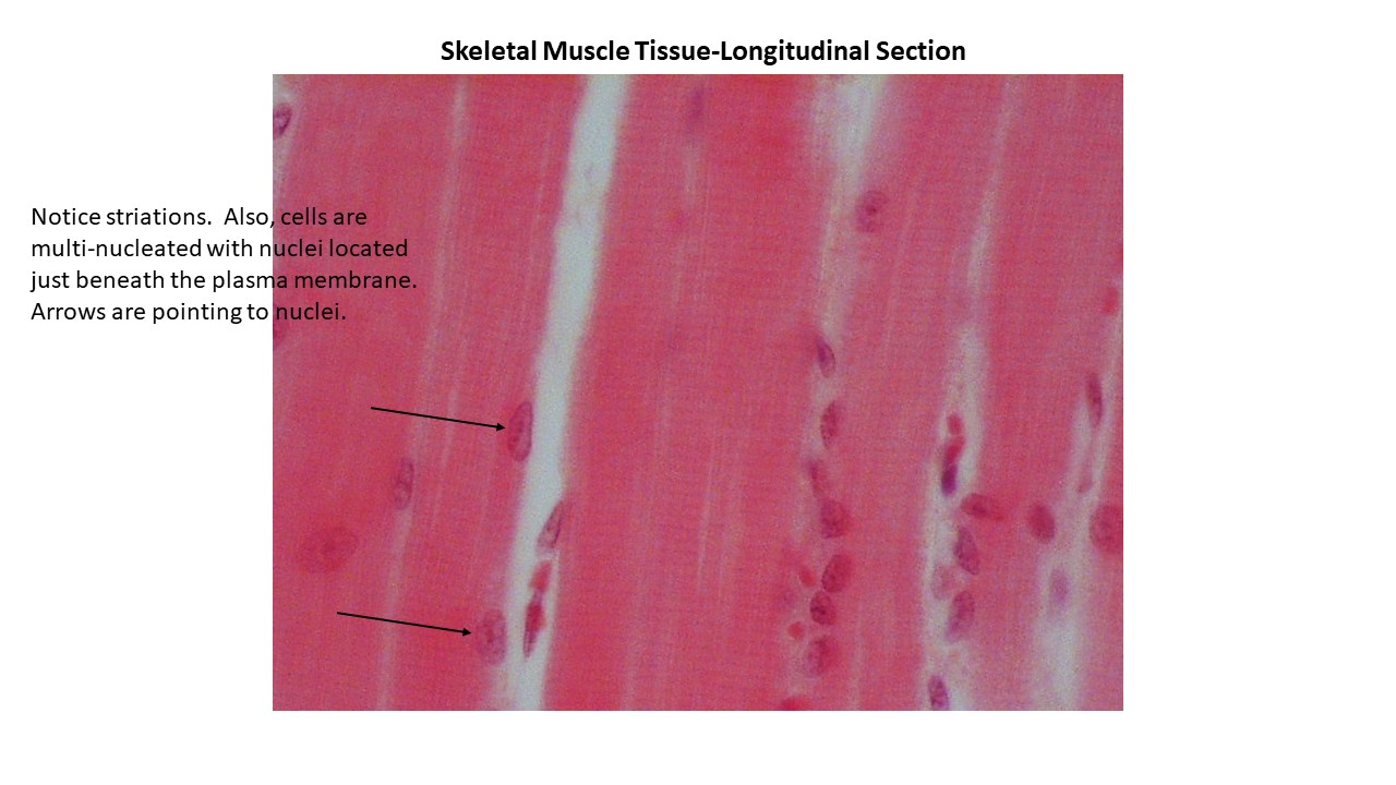
• Skeletal Muscle- Multi-nucleated cells
- Striations
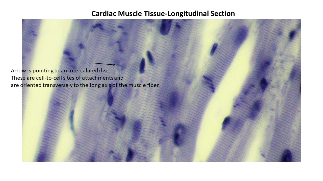
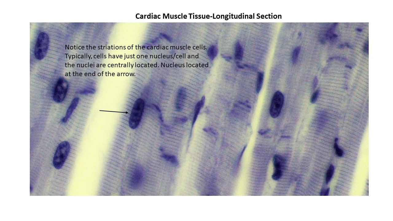
- Cardiac Muscle
- Single, centrally locate nucleus
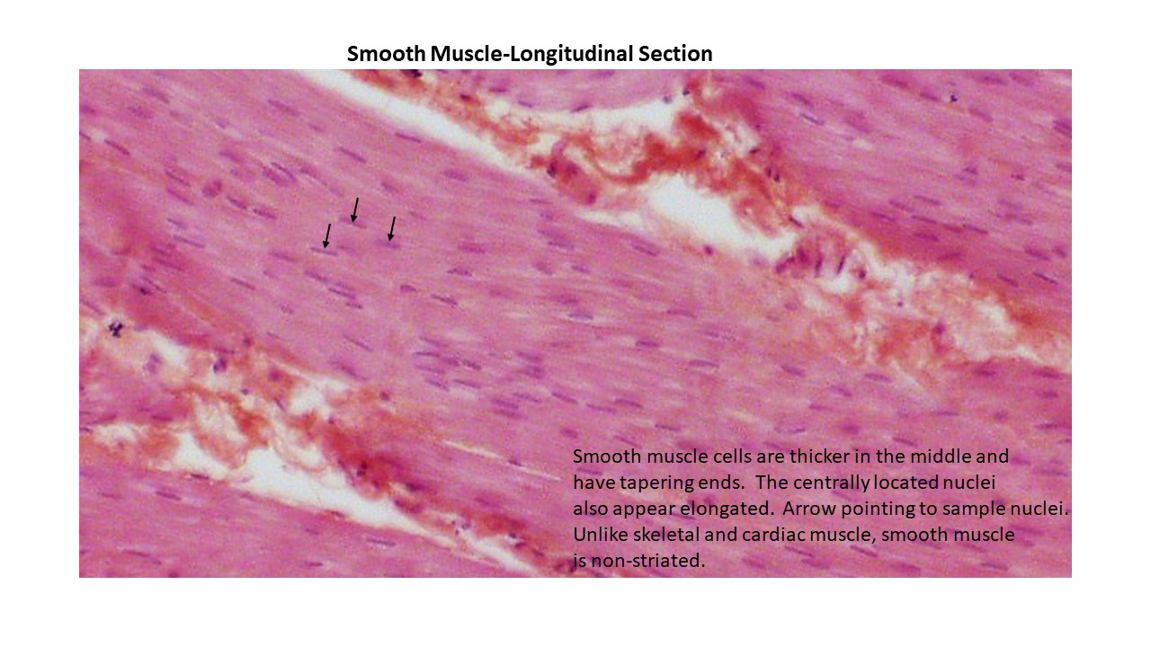
- Smooth Muscle
- Centrally located nucleus
- Non-striated
Histology Slides: Be able to identify the three types of muscle tissue on a light microscope and the labelled structures for each.
- Skeletal Muscle
- Skeletal Muscle
- Long, cylindrical cells
- Locate the nuclei of the multi-nucleated fibers (cells)
- Striations
- Skeletal Muscle
- Cardiac Muscle
- Cardiac Muscle with Intercalated Disc
- Cardiac Muscle Cell with Prominent Striations and Centrally Located Nucleus
- Short-branched cell
- Centrally located nucleus
- Intercalated discs
- Striations
- Smooth Muscle
- Smooth Muscle
- Spindle-shaped cell
- Centrally located nucleus
- Non-striated
- Smooth Muscle
Axial Muscles
- Muscles of Facial Expression
- Frontalis and Occipitalis (aka epicranius, aka frontal belly and occipital belly of the occipitofrontalis)
- Orbicularis Oris
- Zygomaticus Major and Minor
- Buccinator
- Platysma
- Orbicularis Oculi
- Risorius
- Mentalis
- Masseter
- Depressor Anguli Oris
- Depressor Labii Inferioris
- Platysma
Extrinsic Eye Muscles
-
- Superior Rectus
- Medial Rectus
- Lateral Rectus
- Inferior Rectus
- Superior Oblique
- Inferior Oblique
- Muscles of Mastication (chewing muscles)
- Temporalis
- Masseter
- Muscles That Move the Head and Neck
- Sternocleidomastoid
- Muscles of Respiration
- External Intercostals
- Internal Intercostals
- Diaphragm
- Muscles of the Abdominal Wall
- Rectus Abdominis
- External Oblique
- Internal Oblique
- Transversus Abdominis
Appendicular Muscles
- Muscles That Move Pectoral Girdle
- Pectoralis Minor- anterior muscle
- Serratus Anterior- anterior muscle
- Trapezius- posterior muscle
- Muscles That Move the Glenohumeral Joint/Arm
- Pectoralis Major- anterior muscle
- Latissimus Dorsi- posterior muscle
- Deltoid
- Rotator cuff muscles attach the scapula to the humerus
- Supraspinatus
- Infraspinatus
- Subscapularis
- Teres minor
Arm and Forearm Muscles That Move the Elbow Joint/Forearm
- Flexors of Forearm
- Biceps briachii
- Biceps brachii
- Brachialis
- Brachioradialis
- Forearm Extensors
- Triceps Brachii
Muscles of the Pelvic Girdle and Lower Limb
- Muscles That Move the Hip Joint/Thigh
- Adductor Magnus- adducts and flexes thigh
- Psoas Major
- Iliopsoas
- Thigh Muscles That Move the Knee Joint/Leg
- Quadriceps Femoris- Leg Extensors
- Rectus Femoris
- Vastus Lateralis
- Vastus Medialis
- Vastus Intermedius
- Sartorius- leg flexor, medial rotation of leg
- Gracilis- flexes and adducts thigh, leg flexor
- Gluteus Maximus- extends thigh, lateral rotation of thigh
- Gluteus Medius-abducts thigh, medial rotation of thigh
- Hamstrings Group- posteriorly thigh muscles; extensors of thigh, leg flexors
- Semimembranosus-Medial
- Semitendinosus-Medial
- Biceps Femoris-Lateral
- Muscles of Leg and Foot-Posterior, Anterior and Lateral Compartments
- Gastrocnemius- posterior compartment; flexes leg, plantar flexion of foot
- Soleus- posterior compartment
- Tibialis Anterior- anterior compartment; dorsiflexion of foot
- Fibularis Longus (or peroneus longus)- lateral compartment; everts foot, plantar flexor