
General clinical signs observed in the skin
A. Jaundice
- In individuals with lighter skin tones, jaundice presents as yellowing of the skin, eyes, and mucous membranes.
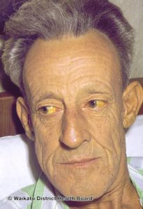
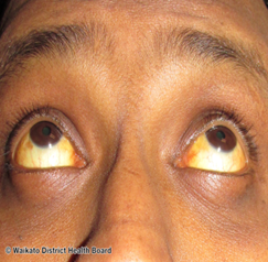
Clinical Presentation of jaundice in an individual with Fitzpatrick Skin Type II DermNetNZ.org - In individuals with darker skin tones, jaundice is not as evident on the skin. However, it may be detected in oral mucosa or the sclera or white of the eyes.
B. Cyanosis
- In individuals with lighter skin tones, cyanosis appears as a bluish discoloration of the lips and nail beds. In individuals with darker skin tones, cyanosis presents as a grayish discoloration along the buccal mucosa.
C. Inflammation
- The cardinal signs of inflammation include erythema, edema, increased temperature, and pain. Erythema can present differently in patients of varied skin tones. Erythema is lighter and mid-skin tone patients will present as redness.
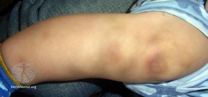
D. Bruising
- Bruising occurs when capillaries under the skin are damaged. The superficial skin remains intact.
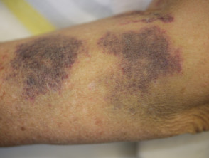
- In patients with a lighter skin tone, bruises present initially as a purple, red, or black discoloration. As time progresses, the bruising may change to a yellowish/greenish color to lighter brown/yellow color.
Bruising caused be hemophilia on an individual with Fitzpatrick Skin Type III DermNetNZ.org - In individuals with darker skin tones, bruising presents more as a dark purple or black discoloration.

Traumatic bruise on a individual with Fitzpatrick Skin Type IV DermNetNZ.org
E. Pallor
- In individuals with lighter skin tones, pallor is seen as a pale color in the skin or nail beds. In darker skin tones, pallor may look ashen or gray, while in brown toned skin it can appear yellow. However, in persons with darker skin tones the palmar surface of the hands and feet may still appear pale.
F. Dehydration
- Dehydration causes skin changes such as gray color in lighter skin tones. In darker skinned individuals, hyperpigmentation or darker shadows may be visible around the eyes. In all skin tones fine lines may also be visible.
G. Dry skin
- Dry skin can present as skin that is rough, scaly, or cracking.

H. Urticaria (hives)
- Urticaria presents as wheals (elevated patches) that may appear redder or paler than surrounding skin in patients with a lighter skin tone. In a patient with a darker skin tone the hives may just present as elevated patches that are slightly darker than than the natural skin tone. Patients may also have itching around the area.

I. Hyperpigmentation
- Abnormally increased pigmentation, due to increased melanin production. Temporary hyperpigmentation can occur post-injury or disease. It is prevalent in all skin tones, but occurs more commonly in individuals with a darker skin tone.

J. Purpura
- Bleeding that results in collection under the tissue in larger flat areas. that are a reddish-purple patch. The regions are about 4 mm to 1 inches.

K. Ecchymosis
- A very large bruised area. This can occur when multiple blood vessels close together rupture. This results in the pooling of blood just under the skin surface. It looks like a bruise, but is not necessarily related to an injury.

Endocrine & Metabolic Conditions
A. Hyperpituitarism – Cushing Disease
- Cushing Disease: A common form of hyperpituitarism closely related, but distinct from, Cushing Syndrome. The clinical presentation for Cushing Syndrome (oversecretion of ACTH in adrenal glands) and Cushing Disease (oversecretion of ACTH in pituitary gland) are similar and can include ecchymosis (discoloration of the skin usually caused by bruising) and purple or dark striations on the skin of the trunk abdomen, axillary region, or legs.
B. Hyperthyroidism – Grave’s disease
- Hyperthyroidism: a condition caused by excessive amounts of secreted thyroid hormone. Symptoms of hyperthyroidism include flushed skin due to capillary dilation, and a hard purple or dark colored area on the anterior surface of the tibia that can be accompanied with erythema, pain, or itching. The most common form of hyperthyroidism is Graves Disease. Graves Disease is additionally accompanied by exophthalmos (protruding eyeball) or the presence of a goiter.
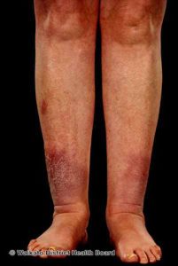
Pretibial Myxoedema in an individual with Graves’ Disease with Fitzpatrick Skin Type III DermNetNZ.org
C. Hypothyroidism
- Hypothyroidism: Also called thyroid hormone deficiency, can be characterized by periorbital and peripheral myxedema (swollen skin with a waxy appearance), dry or flaky skin, and carotenemia (increased levels of beta-carotene levels in the blood).

Carotenoderma in an individual with hypothyroidism, with Fitzpatrick Skin Type II (Left)
DermNetNZ.org
D. Hypoparathyroidism
- Hypoparathyroidism: Caused by a reduction in the secretion of PTH. Integumentary symptoms associated with reduced PTH include skin that is dry, scaly, or coarse. Less commonly, pellagra-like darkened skin can be seen (https://www.ncbi.nlm.nih.gov/pmc/articles/PMC3475912/).
E. Hyperparathyroidism
- Hyperparathyroidism– Can lead to bone decalcification and hypercalcemia in the blood. Calcinosis cutis (calcium deposits in or beneath the skin) or bone cysts can deposit and push up against the skin, leading to a pale discoloration that is hard on palpation.
Calcinosis Cutis on an individual with Fitzpatrick Skin Type II DermNetNZ.org
F. Primary adrenal insufficiency – Addison disease
- Addison Disease: Also called Primary Adrenal Insufficiency, Addison Disease increases the prevalence of melanocyte-stimulating hormone which causes increased skin and mucous membrane pigmentation. In darker skinned individuals, this presents as a slate-gray color. In lighter skinned individuals, the skin tone can range from a slight local tan to a global bronze-like tan.
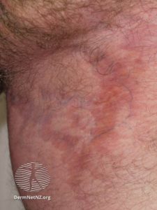
G. Adrenocortical hyperfunction – Cushing syndrome
- Cushing Syndrome: A condition that results from excessive cortisol secretion. Symptoms present similarly to Cushing Disease and can include increased ecchymosis due to excess protein tissue wasting, a characteristic moon shaped face, and a thinning of skin with excessive purple or dark colored striae.
Striations in the Axillary region of an individual with a Fitzpatrick Skin Type II DermNetNZ.org
H. Diabetes mellitus
- One common complication of diabetes mellitus (DM) is diabetic foot ulcers.1
- Patients with DM can also present with dry, inelastic skin due to loss of autonomic nerve function. In later stages of DM, atrophic changes continue. The skin thins out and becomes shiny, cyanotic, and dry.1
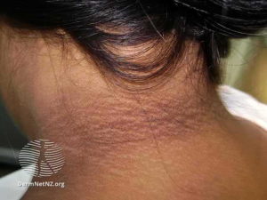
- Other skin conditions that may present in patient’s with DM:
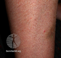
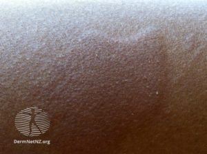
- Scleroderma diabeticorum is an asymptomatic thickening of the skin. Most commonly in the posterior neck, upper back, and shoulders. Scleroderma diabeticorum can lead to erythema, hyperpigmentation, or an orange peel appearance of the skin (Peau d’Orange).1
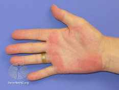
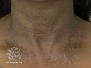
Postinflammatory hyperpigmentation on an individual with Fitzpatrick Skin Type II DermNetNZ.org Acquired dermal macular hyperpigmentation on an individual Fitzpatrick Skin Type V DermNetNZ.org Palmar erythema on an individual with Fitzpatrick Skin Type II DermNetNZ.org - Acanthosis Nigricans (AN) is a condition causing velvety, dark patches of the skin in areas of the body such as the elbows or knees. 2
Acanthosis nigricans presentation on an individual with Fitzpatrick Skin Type V. DermNetNZ.org 
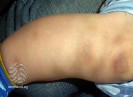
Acanthosis nigricans presentation on an individual with Fitzpatrick Skin Type II. DermNetNZ.org - Diabetic Dermopathy/Shin Spots are red or brown round patches lining the skin up the front of the legs.2 In patients with darker skin tones, the spots present as more brown whereas in lighter skin tones it presents as red.
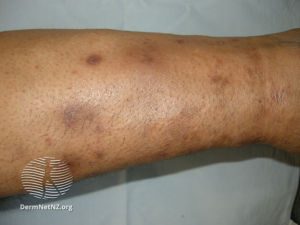
Shin spots presenting on an individual with Fitzpatrick Skin Type V, as evidenced by the brown color of the spots. DermNetNZ.org 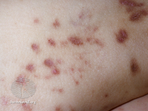
Shin spots presenting on an individual with Fitzpatrick Skin Type I, as evidenced by the reddish hue of the spots. DermNetNZ.org - Necrobiosis Lipoidica is a condition causing yellow, red, or brown patches on the skin. The areas start as small, raised, pimple-like bumps that progress into hard, swollen patches.2
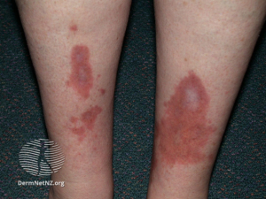
Presentation of necrobiosis lipoidica in an individual with Fitzpatrick Skin Type II. DermNetNZ.org - Bullosis Diabeticorum/Diabetic Blisters/Diabetic Bullae is a condition that appears like burn blisters that develop on the lower legs and feet. These should look similar in patients of all skin tones.2

Diabetic blisters presenting in an individual with Fitzpatrick Skin Type VI. DermNetNZ.org 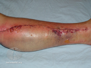
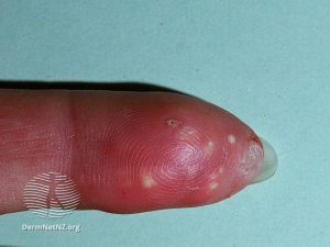
Diabetic blisters presenting in a patient with Fitzpatrick Skin Type III. DermNetNZ.org - Eruptive Xanthomatosis is a condition causing small bumps on the back of hands, feet, arms, legs, and buttocks that present as reddish-yellow.2
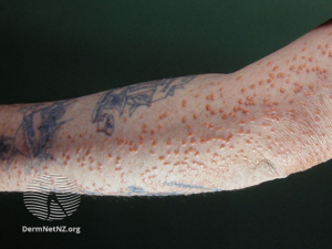
An individual with Fitzpatrick Skin Type I, presenting with eruptive xanthomatosis. DermNetNZ.org 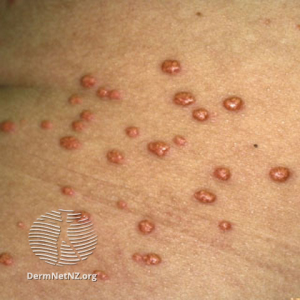
Close up view of eruptive xanthomatosis in an individual with Fitzpatrick Skin Type IV. DermNetNZ.org - Digital Sclerosis begins with tight, thick waxy skin on the fingers that can progress to hard, thick, swollen skin spread throughout the body.2
- Carotenodermia– yellowish skin and nails.3
Carotenoderma in an individual with hypothyroidism, with Fitzpatrick Skin Type II (Left)
DermNetNZ.org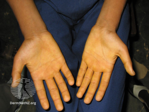
Carotenoderma presenting in an individual with Fitzpatrick Skin Type V. DermNetNZ.org - Vitiligo is a loss of skin pigmentation in a spotted manner. It appears in all skin tones as splotches of skin that are lighter in tone than the rest of the body.3
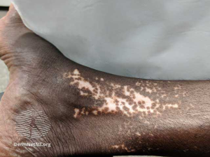
An individual with Fitzpatrick Skin Type VI, presenting with vitiligo. DermNetNZ.org 
An individual with Fitzpatrick Skin Type III, presenting vitiligo. DermNetNZ.org - Bacterial Infections are common in patient’s with diabetes mellitus. These present as hot, swollen, red, painful skin, hair follicles, or fingernails.2
- Fungal Infections are also common in patients with diabetes mellitus. These present as itchy, red rashes surrounded by tiny red blisters and scales.2
- Scleroderma diabeticorum is an asymptomatic thickening of the skin. Most commonly in the posterior neck, upper back, and shoulders. Scleroderma diabeticorum can lead to erythema, hyperpigmentation, or an orange peel appearance of the skin (Peau d’Orange).1
I. Wilson’s disease/Hepatolenticular Degeneration
- One common symptom of chronic Hepatolenticular Degeneration is liver disease resulting in jaundice. In individuals with lighter skin tones, jaundice presents as yellowing of the skin, eyes, and mucous membranes. In individuals with darker skin tones, jaundice is not as evident on the skin. However, it may be detected in oral mucosa or the eyes.
- It can also cause Fleischer rings around the iris of the eye due to copper deposits. These copper deposits in the eyes should look similar in people of all skin tones.
- This disease can also cause degenerative changes in the brain, particularly the basal ganglia, which may require physical therapy rehabilitation.

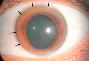
Low QJ, Siaw C, Lee RA, Cheo SW. Kayser-Fleischer rings and Wilson’s disease.
References
- Goodman & Fuller’s Pathology: Implications for the Physical Therapist. 5th ed. 2020.
- https://www.cdc.gov/diabetes/library/features/diabetes-and-your-skin.html
- Cutaneous Manifestations of Diabetes Mellitus: A Review. Am J Clin Dermatol. 2017 Aug;18(4):541-553. doi: 10.1007/s40257-017-0275-z.
- Mucocutaneous manifestations of acquired hypoparathyroidism: An observational study
