
Cardiopulmonary Assessment
A. Skin appearance
i. Pallor (paleness)1
- Pallor is characterized by a pale appearance of skin, nail beds, and mucus membranes. In individuals with darker skin tones, pallor may present as ashy gray. In individuals with brown skin tones, it may appear more yellowish.
- To help identify pallor in individuals with darker skin tones, one may assess the palms or the sole of the feet, which should appear paler if pallor is present.
ii. Cyanosis
- In individuals with lighter skin tones, cyanosis appears as a bluish discoloration of the lips and nail beds. In individuals with darker skin tones, cyanosis presents a grayish discoloration along the buccal mucosa.2 Therefore an inspection of the inside of a patient’s mouth may be necessary.
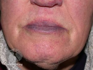
Cyanotic lips on an individual with Fitzpatrick Skin Type II DermNetNZ.org.
iii. Edema
- A condition in which there is an excessive accumulation of interstitial fluid, resulting in swelling. Edema should present similarly in all types of skin tones.2
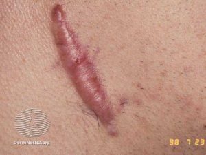
iv. Scarring
- Individuals with darker skin tones may present with hypopigmentation at the area of scarring, resulting in increased susceptibility to sunburn. Additionally patients with darker skin tones are more prone to keloid scarring.

B. Neck
i. Jugular venous distension
- Jugular venous distention (JVD) is a possible sign of right-sided heart failure. Fluid overload results in increased fluid retention, causing fluid to be backed up into the lungs and venous system, resulting in a swollen jugular vein. JVD should present similarly amongst different skin tones.
C. Extremities
i. Nicotine stains
- Nicotine turns yellow when oxidized, resulting in discoloration. This discoloration is present on skin, teeth, and fingernails.3
- There is currently not much research conducted on nicotine staining amongst different skin tones. To account for possible differences in nicotine staining on different skin tones, look at nails and teeth for presence of discoloration.

ii. Vascular lesions
- Vascular lesions differ in appearance and are caused by a variety of factors, including capillary malformation or trauma. Due to the varied nature of their appearance, it is important to determine the origin of the lesion.
- Examples of vascular lesions:
- Trauma (e.g. nail bed hemorrhages)
- Vasculitis: blood vessel inflammation
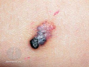
- Cherry angioma: a benign lesion that presents as a small, raised papule. The papule may be red, blue, or purple in color. It may also appear black, when thrombosed.
- Examples of vascular lesions:

-
-
- Angiokeratoma: hyperkeratosis that results from capillaries dilating or rupture in the superficial dermis. The papule may appear blue, red, or black depending on the stage of healing.
-

D. Vital signs
i. Oxygen saturation
- Oxygen saturation measurements can be less accurate in patients who identify as black. This is due to a higher prevalence of occult hypoxemia, which is when an arterial oxygen saturation of <88% despite an oxygen
saturation of 92 to 96% on pulse oximetry. In a large study, occult hypoxemia is present in 11.4% of black patients compared to 3.6% of white patients. - The variation in occult hypoxemia risk according to race necessitates the integration of pulse oximetry with other clinical measures.4
E. Special tests
i. Capillary refill test
- The capillary refill test is a measure of arterial flow at the skin surface. The test requires an examiner to compress the nail bed and hold for 5 seconds which will cause blanching, upon release the blanched area should return to the previous color in 3 seconds or less to be considered normal. However, the accuracy of this test can be influenced by multiple factors including skin color and the lighting in the room.
- In patients with a lighter skin tone who have nail polish or artificial nails it is recommended to perform on the adjacent skin area, however this is not possible in patient’s with a darker skin tone since the melanin will prevent the ability to blanch the skin.
- A prolonged capillary refill time can indicate arterial insufficiency5
ii. Elevation pallor
- An elevation pallor test assesses arterial perfusion. The limb is elevated 60 degrees and held in position for 60 seconds, during elevation the sole of the foot is observed for pallor. A normal test result would be no pallor and an abnormal test result is the onset of pallor, which can be timed and a grade assigned. Pallor will present differently based on skin tone.
- In lighter skin tones, the pallor will appear as very pale pink, peach or white.
- In brown skin tones pallor will appear yellow-brown.
- In dark brown or black skin tones, pallor will appear an ash gray.
iii. Dependent rubor
- The dependent rubor test is a test for arterial insufficiency, after elevation of the limb at 60 degrees for 60 seconds, the limb is moved to a dependent position and observed for color changes.
- In patients with lighter skin tones, the rubor will present as a red or pink color.
- In patients with darker skin tones, the rubor will present as either a dark purple color or as a shade darker than their normal skin tone.
iv. Venous filling time
- The venous filling time test is a test for vascular insufficiency. The lower extremity is elevated, until the superficial veins on the dorsal of the foot are drained by gravity. Afterwards, the limb is moved to a dependent position and the foot is observed for the speed at which the superficial veins refill with blood.
- In patients with darker skin tones, venous drainage and refill is not as evident. Palpation may be a better alternative, to determine the rate of venous drainage/refill.
References
- https://mvec.mcri.edu.au/references/identifying-aefi-in-diverse-skin-colour/
- Goodman CC, Fuller KS. Pathology: Implications for the Physical Therapist. 5th ed. 2021.
- https://www.sciencedirect.com/science/article/pii/S1570023220301707
- Sjoding MW, Dickson RP, Iwashyna TJ, Gay SE, Valley TS. Racial bias in pulse oximetry measurement. N Engl J Med. 2020:383;25.
- https://dermnetnz.org/topics/arterial-ulcer
