
Integumentary conditions
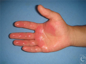
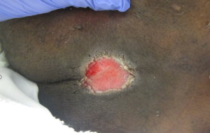
A. Cellulitis
An active bacterial infection that occurs under the surface of the skin. Cellulitis will cause bright redness and swelling in lighter skin tones. In darker skin tones, it can appear swollen, red, purple, or darker than the surrounding tissues.


B. Lymphedema
A chronic condition that is caused by lymphatic insufficiency. It can be caused by genetic malformation or acquired injury to the lymphatic system.
- Clinical signs present include edema and skin changes that will vary depending on the stage of the lymphedema.
- Stage 1 lymphedema will present with non-pitting edema that can resolve with elevation. Edema will present similarly in all Fitzpatrick skin types.
- Stage 2 lymphedema clinical signs include pitting edema and fibrosis of the skin. Fibrosis is a hard texture based on palpation that will have a different appearance in Fitzpatrick skin types.
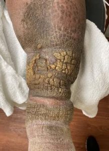
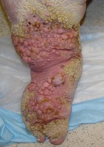
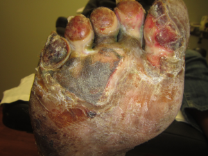
- Stage 3 lymphedema will present with significant skin fibrosis, hyperkeratosis, peau d’orange, elephantiasis, and papillomas. In this stage of lymphedema, many of the clinical signs will present differently depending on the Fitzgerald skin type.
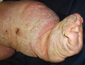
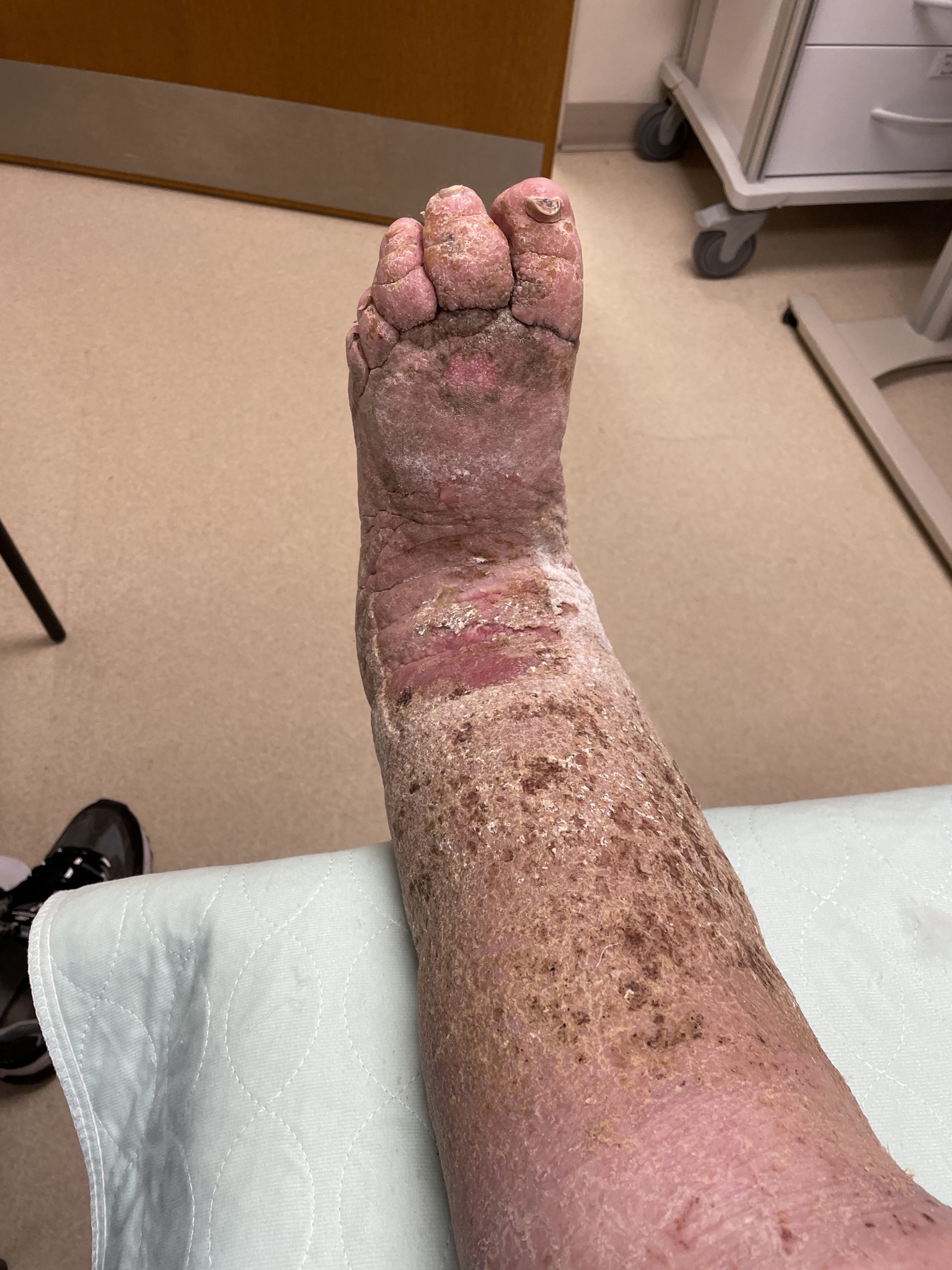
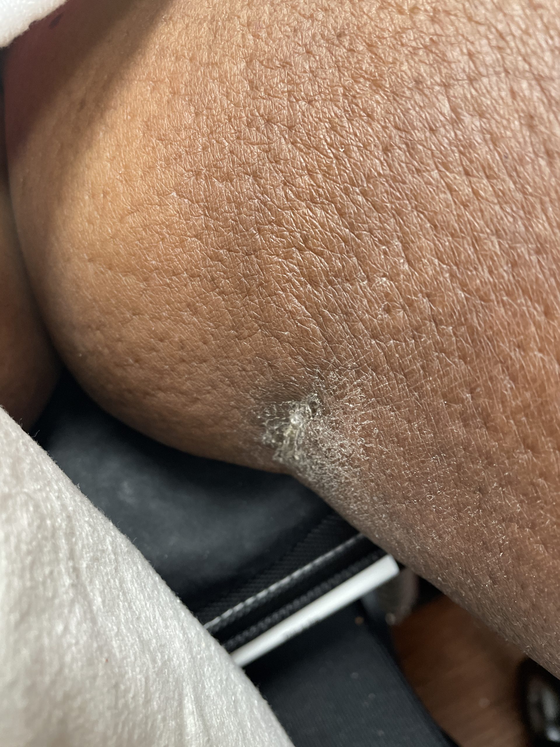
Hyperkeratosis is an over production of epithelial tissue that creates thick plaques. Plaques will look similar in different Fitzpatrick skin types, with either a brown of black discoloration.
Peau d’orange has a distinctive orange peel skin texture with enlarged pores. In lighter skin tones enlarged pores will be a distinct pink, purple, or red. In darker skin tones enlarged pores will appear dark purple or black.

Papillomas are highly vascular skin tags caused within the final stages of lymphedema. Papillomas usually present flesh-colored or pink for all Fitzpatrick skin types.
C. Oncology
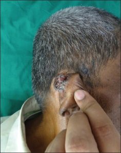
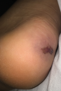
Basal cell carcinoma
- The most common form of skin cancer which develops from the uncontrolled growth of basal cells. Sun exposure is the most common cause, and individuals with lighter skin tones are more likely to develop basal cell carcinoma (BCC) than individuals with darker skin tones.
- It appears similarly in all skin tones as a slightly elevated red or pink mark with rolled edges that and can include surface level widened blood vessels (telangiectasia).4
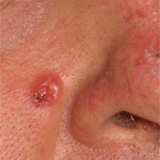
BCC in Fitzpatrick skin type I.
Squamous cell carcinoma
- The most common type of malignant skin cancer in darker skin tones and the second most common type of malignant skin cancer in lighter skin tones. SCC affects the keratinocytes in the epidermis. It is more common in light skinned individuals. It mostly affects the epidermis but can metastasize deeper into the dermis. In lighter skin tones it may present as flesh colored or red, with surrounding tissues appearing scaly or thickened. In darker skin tones, a scaly or hyperkeratotic and pigmented plaque is common.4

Melanoma
- Superficial spreading melanoma is the most common form of melanoma. It typically presents similarly in all skin tones, but in darker skin tones it can present with thicker or more advanced tumors as a result of increased disease progression due to decreased rates of detection. Melanoma tumors can manifest as a raised patch of brown or black with irregular borders, a large diameter, uneven coloration, and asymmetric shape. They also change in size, shape, or color.4

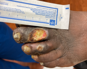
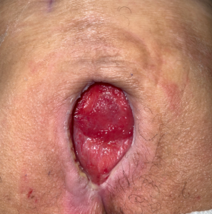
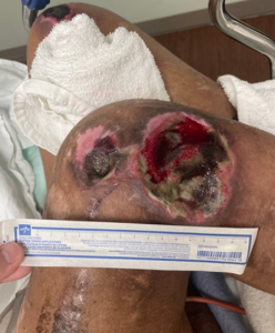
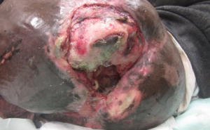
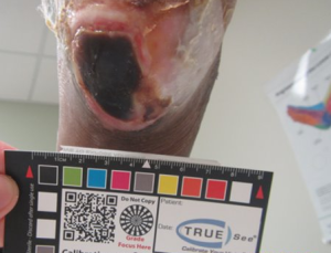
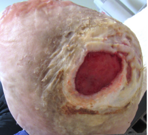
Malignant cutaneous wounds/fungating ulcers
- Malignant tumors are an overgrowth of cells outside the margins of the skin. Visual appearance is the same across all skin tones as the tumors are made of granulation, slough, and/or eschar. Tumor margins typically have erythema which will present pink/red in lighter skin tones and purple/maroon in darker skin tones.


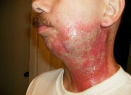
D. Burns
Burns can occur from a series of mechanisms: Thermal (flame, contact, scald, cold), chemical, radiation, and electrical. Classification of the burn is based on the depth of penetration which is similar across all mechanisms. Frostbite (cold) mechanism of injury has some specific differences which are categorized below.6
Superficial injuries (previously known as first degree burns):
- Superficial injuries are characterized by injury to the epidermis only and allow for complete recovery.
- Bright red discoloration in lighter skin tones and maroon or purple in darker skin tones.
- A superficial injury under the thermal mechanism of frostbite is categorized as frostnip. Clinical appearance is the same as other superficial injuries.


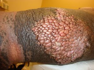
Partial thickness injury (previously known as second degree burn)
- Two categories within the partial thickness injuries: Superficial partial-thickness and deep partial-thickness injuries.
- Superficial partial-thickness injury occurs at the papillary dermis creating serous-filled blisters. Sensation is intact.
- Deep partial-thickness injury occurs deeper within the dermis. Sensation is affected.
- Superficial frostbite can present with clear or serous blisters, hyperemia, and numbness. The skin may present with a blotchy blue-gray color or with erythema.8


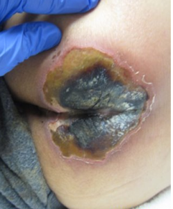
Full thickness injury (previously known as third degree burn)
- Damage to all skin layers and causes permanent tissue damage.
- Normally characterized by black or brown eschar without sensation.
- Deep frostbite becomes white until it thaws and turns into a blue-gray appearance with a firm, woody end feel.


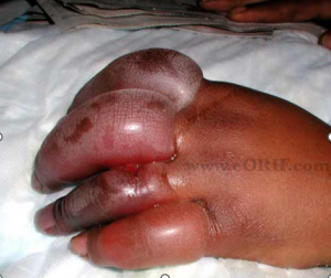
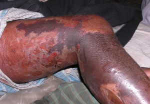
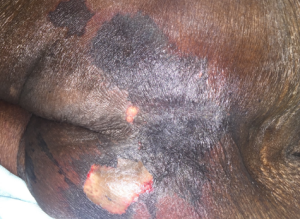
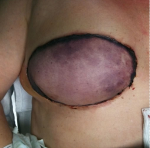
E. Necrotizing fasciitis
- A significant infection of the subcutaneous tissue that progresses rapidly causing significant damage and systemic toxicity if not addressed quickly and appropriately.6
- The affected area will swell and may show a purplish rash. These dark marks can form into blisters with dark fluid. The skin in the area can then start to become dark and necrotic.


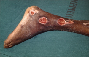
F. Arterial ulcers
- Dry, small, well-defined wounds that contain necrotic tissue secondary to poor arterial flow. This should look similar in patients of all skin tones.
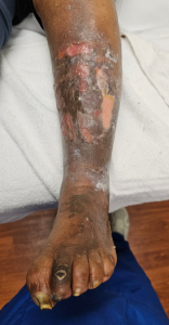
G. Venous ulcers
- Shallow wounds with a granular or fibrotic base and irregular borders. The surrounding skin will present as hairless, pale, & shiny.
- Venous wounds present with characteristic features including non-pitting edema, hemosiderin staining, and atrophie blanche.
Non-Pitting Edema is swelling that does not retain finger impression after release.
Hemosiderin Staining is discoloration due to leakage of red blood cells that migrate to the dermis & epidermis. The skin has a brownish-purple discoloration in lighter skin tones and dark brown to black discoloration in darker skin tones. Hemosiderin staining primarily presents around the distal LE.

Atrophie blanche is blanching of the skin that occurs due to chronic venous insufficiency. The skin presents with a lighter tone relative to the person’s natural color due to decreased blood flow.

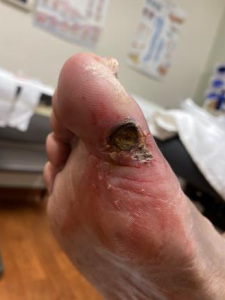
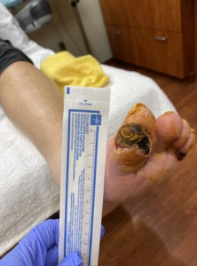
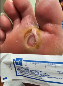
H. Neuropathic ulcers
- These wounds are caused by repetitive trauma to the weight-bearing surfaces of the foot.6 The visual presentation will be similar across all skin types.

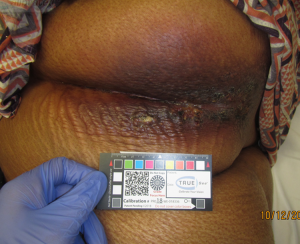
I. Pressure injuries
- Wounds that occur over a bony prominence where there has been an extended time of unrelieved pressure.
- Pressure injuries are classified by Stages I-IV, deep tissue pressure injury, and unstageable.
Stage I: Non-blanching erythema
- This will present as non-blanching red/pink in all skin tones. Can be difficult to assess in patient’s with darker skin tones.


Stage II: Partial-thickness injury
- Similar to superficial burns, these wounds occur at the level of the superficial dermis. These wounds can present as blisters or open wounds with margins at the level of the surrounding skin. There will be no necrotic tissue present. These wounds will present similarly across all skin tones.

Stage III: Full-thickness injury
- These wounds consist of full dermal damage, but no exposure of deeper structures. These wounds will present similarly across all skin tones.
Stage IV: Full-thickness injury with deeper structures exposed
- These wounds have full dermal damage with muscle, bone, tendon, etc exposed. These wounds will present similarly across all skin tones.
Deep-tissue pressure injury
- These wound are caused by intense and/or prolonged pressure plus shear forces.6
- The discoloration will be deep maroon and/or purple discoloration across all skin tones, however, may be more difficult to assess in patient’s with darker skin tones.

Unstageable injury
- The pressure injury is covered in necrotic tissue that prevents full assessment of the extent of tissue damage. These wounds present similarly across all skin types.
- Once the base is fully visualized, a stage can be determined.
Integumentary assessment
A. Periwound Characteristics
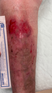
Erythema
- The area encircling the wound that appears redder in lighter skin tones. In darker skin tones, the area can appear darker, purple, or slightly red than the surrounding skin.

Maceration
- Pallor, visible moisture, and wrinkling of the periwound skin due to overhydration and damage of the periwound skin.7

Cyanosis
- This is a sign there is a lack of oxygen to the tissues. In individuals with lighter skin tones, cyanosis appears as a bluish discoloration of the lips and nail beds. In individuals with darker skin tones, cyanosis presents as a grayish discoloration along the buccal mucosa. Once oxygen levels have decreased below metabolic requirements, ischemia occurs. Ischemic tissue is black, dead, and necrotic in all individuals.
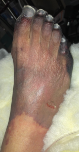

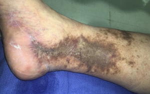
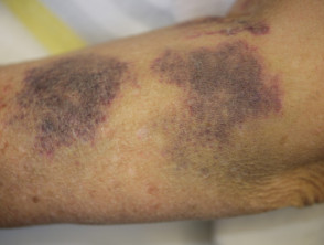
Ecchymosis
- Bruising due to deep tissue trauma. It presents as a dark-red-to-blue discoloration. As it heals, the discoloration can range in colors from bluish-red to green to yellow in person’s with lighter skin tones.6

Venous congestion
- Overperfusion of tissue which can present pink, blue, or dusky. 6

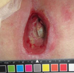
B. Wound bed characteristics
- These wound characteristics will look the same in all types of skin tones: eschar, slough, granulation tissue, muscle, bone, tendon, adipose, wound drainage, rolled edges, uneven edges, tunneling, undermining, fistulas, and sinuses.
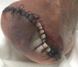
Integumentary interventions
- The wound etiology and periwound/wound bed characteristics are assessed in order to determine the appropriate treatment plan. That plan of care is patient-dependent regardless of skin tone and could include the following:
Debridement
- Selective
- Non-selective
- Enzymatic
Modalities
- Negative Pressure Wound Therapy
- Pulse lavage with suction
- Electrical Stimulation
- Ultrasound – Thermal vs. Non-thermal
- Ultraviolet – C
- Hyperbaric Oxygen Therapy
Compression
- Long vs. short stretch
- Multi-layer compression system
Dressing selection
- Foams
- Hydragels
- Hygrocolloid
- Alginates
- Collagen
References
- Chuangsuwanich A, Chortrakarnkij P, Kangwanpoom J. Cost-effectiveness analysis in comparing alginate silver dressing with silver zinc sulfadiazine cream in the treatment of pressure ulcers. Arch Plast Surg. 2013 Sep; 40(5): 589–596.
- https://www.ncbi.nlm.nih.gov/pmc/articles/PMC3785595/
- https://mvec.mcri.edu.au/references/identifying-aefi-in-diverse-skin-colour/
- https://www.sciencedirect.com/science/article/pii/S0190962205027301?via%3Dihub
- https://www.cdc.gov/ncbddd/jaundice/facts.html
- Hamm, Rose. Text and Atlas of Wound Diagnosis and Treatment. McGraw-Hill Education. 2 ed. 2019.
- Gray M, Bohacek L, Weir D, Zdanuk J. Moisture versus pressure: making sense our of perineal wounds. J Wound Ostomy Continence Nusrs. 2007;34(2):134-142
- Goodman and Fuller. Pathology: Implications for the Physical Therapist. Elsevier. 5th ed. 2020. Page 430.
