
Adverse Effects of Therapeutic Interventions
A. Instrument Assisted Soft Tissue Mobilization (IASTM)
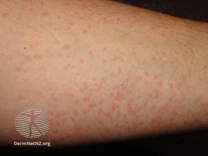
IASTM is a soft tissue mobilization technique that utilizes instruments made from metal, plastic, or a composite material to treat soft tissue restrictions. The technique is applied with enough pressure to disrupt collagen cross links and initiate an inflammatory response in the area of treatment. When the technique is applied, petechiae may start to appear. Petechiae are an indication of small capillary walls being disrupted causing bleeding into the skin. In an individual with a lighter skin tone this will appear as red to purple small spots.1
The appearance of petechiae is an indication that an inflammatory response has been triggered and the area has had enough treatment for that session. If further IASTM is performed over an area of petechiae, it is likely that the area will be over-treated resulting in increased post-treatment pain. In a person with a darker skin tone petechiae are very difficult to visualize since red and purple discoloration is masked by the individuals natural skin tone, therefore other clinical signs of a inflammatory response must be used such as tissue temperature change.
The use of IASTM can result in targeted bruising at the site of the soft tissue restrictions 1-2 days after treatment. On all skin tones bruises will appear darker than the surrounding skin. In an individual with a dark skin tone the bruise will most likely appear a darker brown or even black in the area of the bruise. In an individual with a lighter skin tone, the bruise will initially present as red to purple.
A. 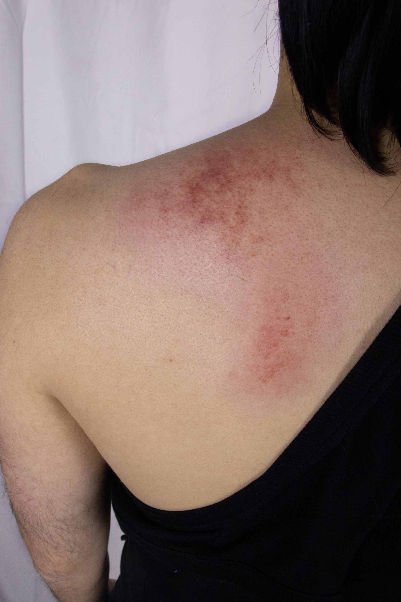 |
B. 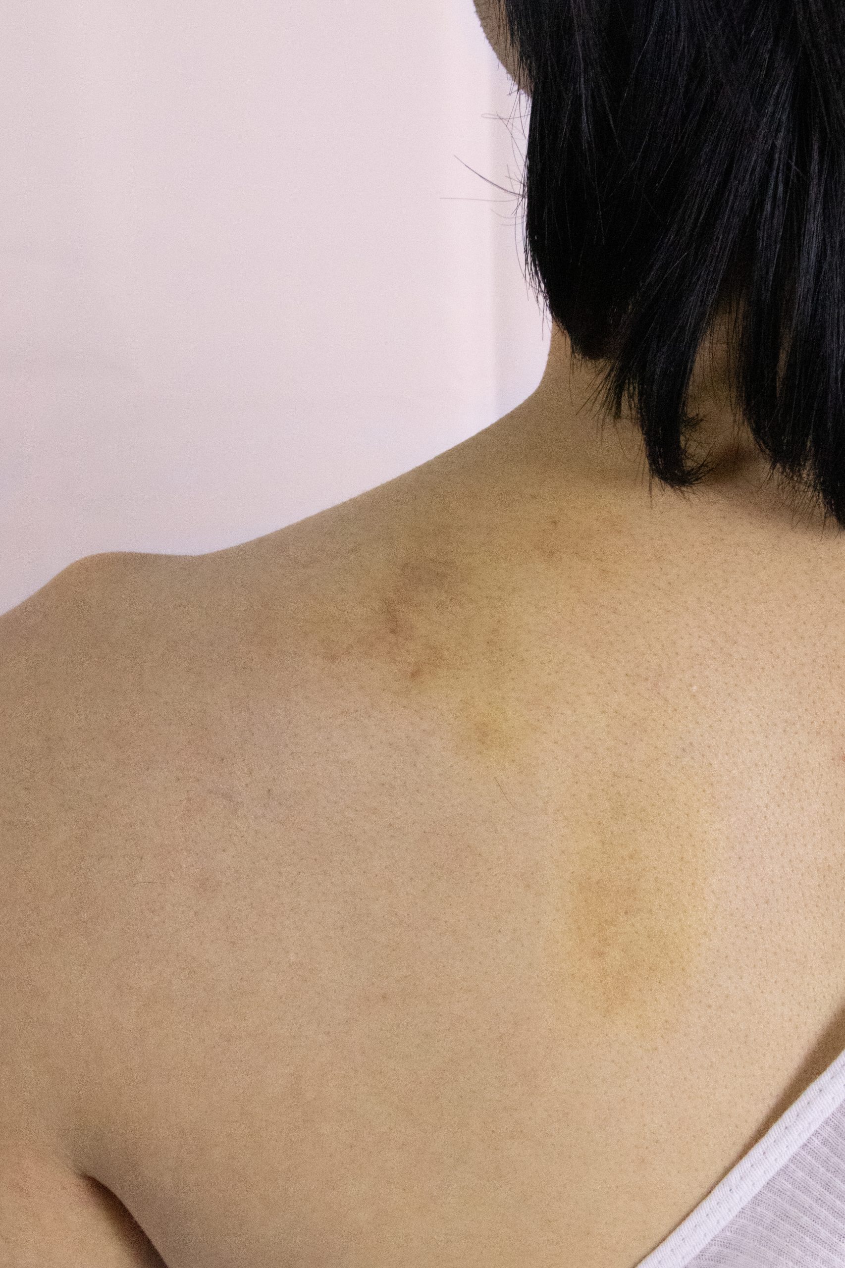 |
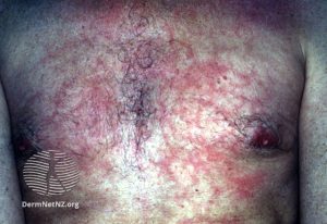
| Figure 1: Pictures A. and B. depict a person with Fitzpatrick skin type III.
Image A was taken just minutes after IASTM was performed on the left upper & middle trapezius muscle. The image shows redness and petechiae due to the modality. Image B. was taken 3 days following the initial IASTM treatment. This image shows yellow-brown bruising from over treatment. |
|
A. 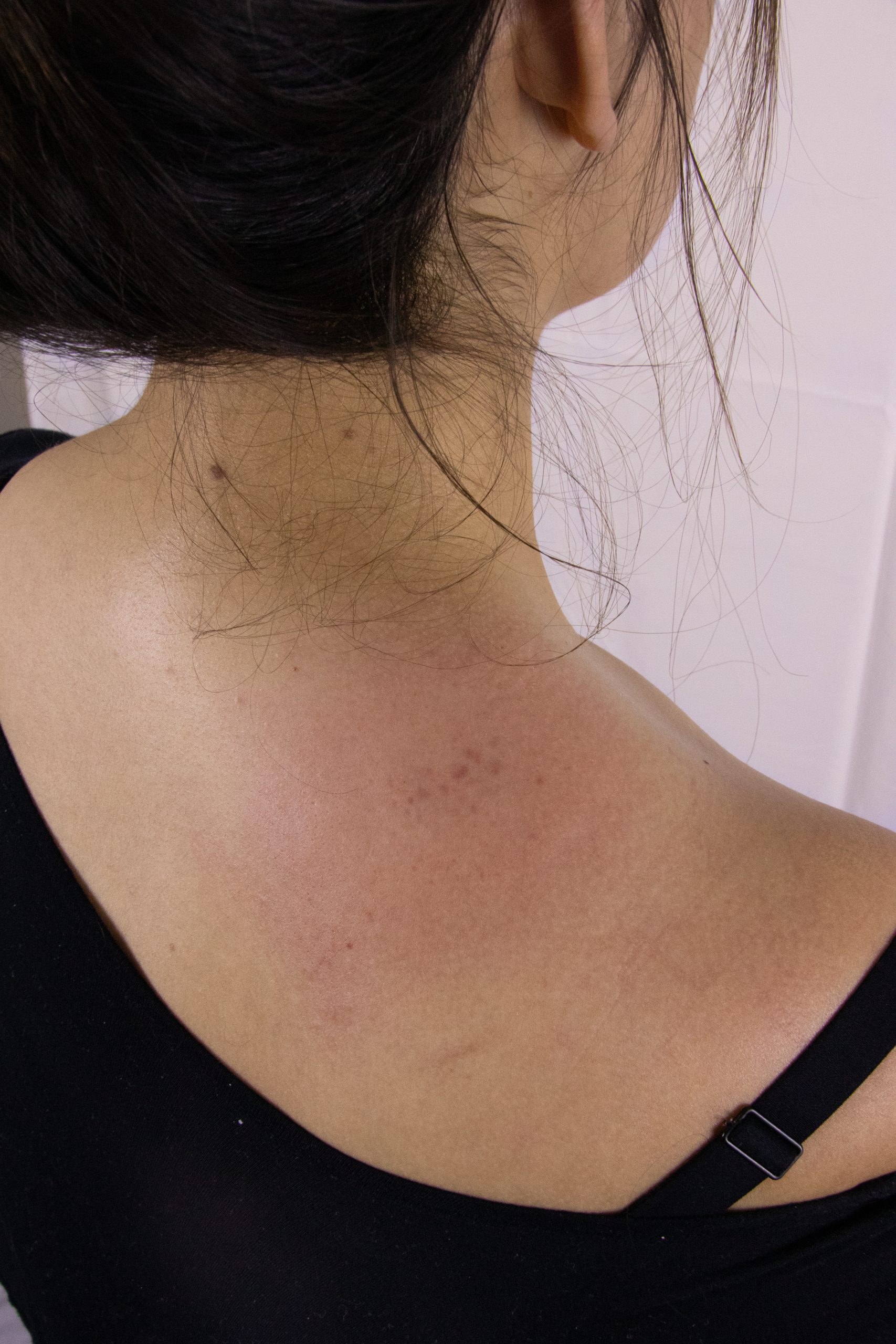 |
B.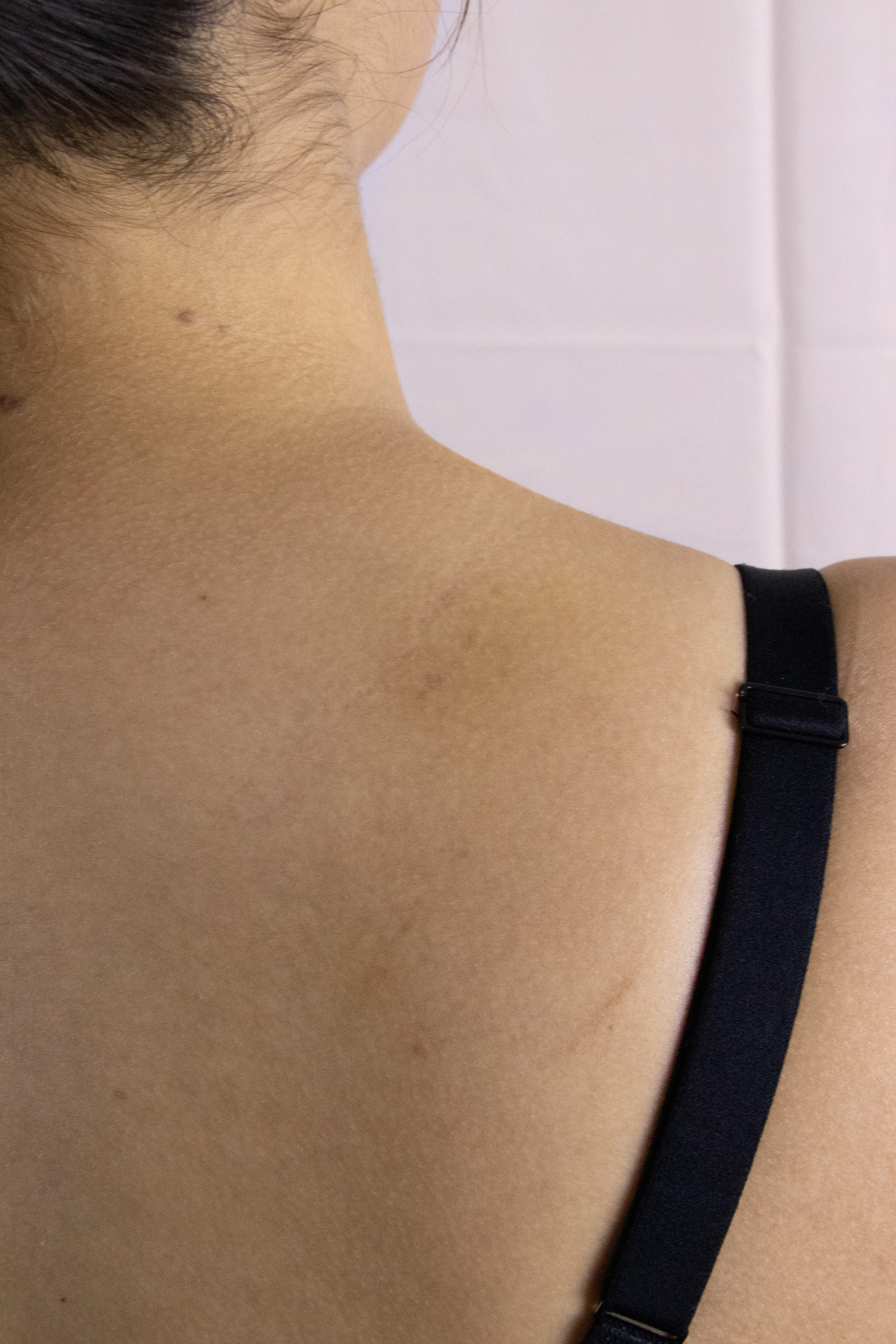 |
| Figure 2: Pictures A. and B. depict a person with Fitzpatrick skin IV.
Image A was taken just minutes after IASTM was performed on the right upper trapezius muscle. The image shows redness and petechiae due to the modality. Image B. was taken 3 days following the initial IASTM treatment. It shows minimal bruising in the area that is a yellow-brown color. |
|
A. 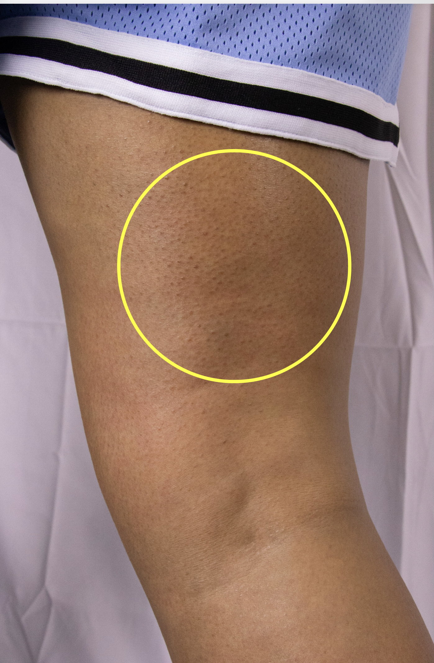 |
B. 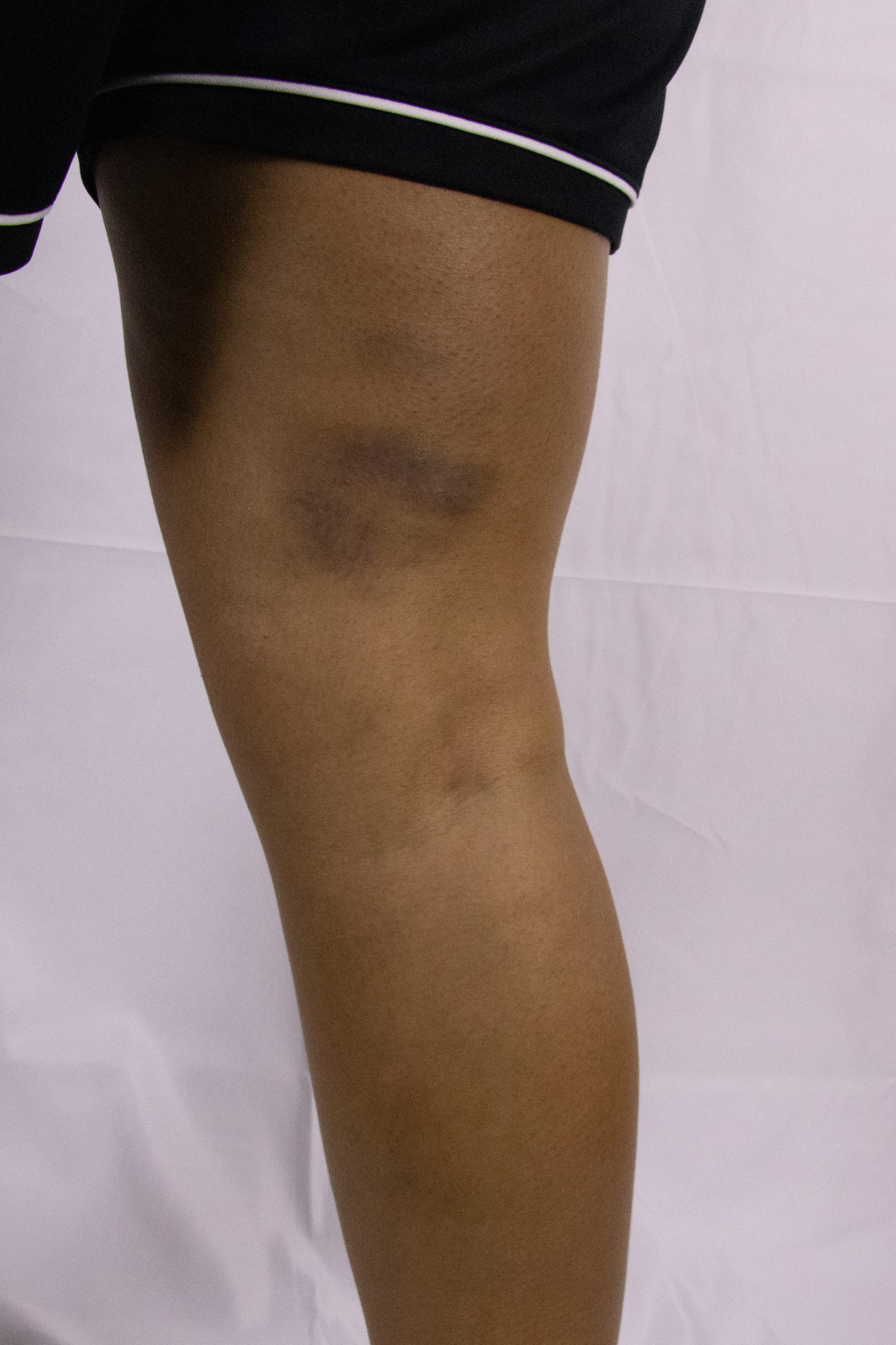 |
C. 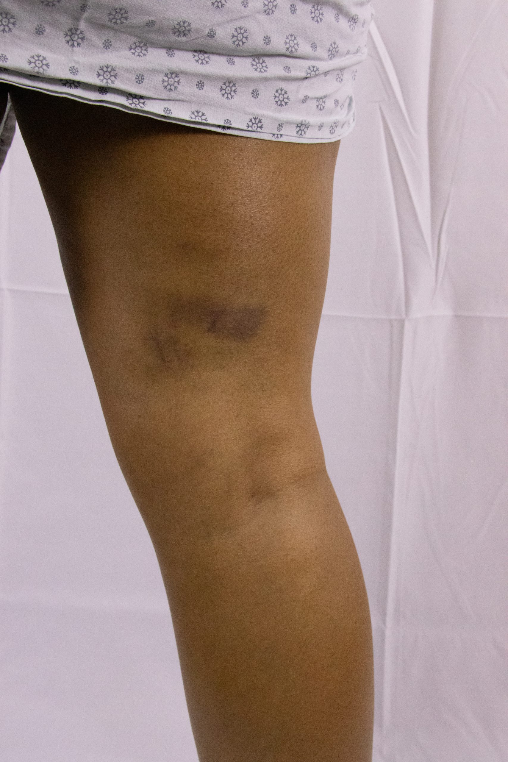 |
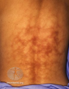
| Figure 3: Pictures A. and B. depict a person with Fitzpatrick sign type V.
Image A was taken just minutes after IASTM was performed on the right hamstrings and gastrocnemius muscles. The image shows a lack of petechiae despite aggressive IASTM to an area of restriction. Other inflammatory signs, such as increased temperature of the skin must be used in patients with darker skin tone to prevent over treatment. Image B. was taken 3 days following the initial IASTM treatment. It shows purple-brown bruising on the medial hamstrings and gastrocnemius muscles due to over treatment with IASTM. Image C. was taken 5 days following the initial IASTM treatment. It shows the progression of bruising as the color lightens. |
||
A.  |
B.  |
| Figure 4: Pictures A. and B. depict a person with Fitzpatrick skin type VI.
Image A was taken just minutes after IASTM was performed on the left middle trapezius muscle. The image shows minimal discoloration and no obvious petechiae, the area of soft tissue that had a restriction was raised and warm to touch. Image B. was taken 3 days following the initial IASTM treatment showed no skin discoloration but some tenderness to touch. |
|
B. Thermotherapy – Hot pack
The most common adverse effect from hot pack application is erythema ab igne. Erythema ab igne is an uncommon condition manifesting in a reticulated, or fishnet-like, pattern of hyperpigmentation on the skin resulting from chronic exposure to low-levels of heat such as a heating pad.2 In a patient with a lighter skin tone the pattern of hyperpigmentation will appear as a red color.
In an individual with a darker skin tone the appearance will be a darker tone of the natural skin tone with an underlying red discoloration.
Another adverse effect of thermotherapy can be a thermal burn which will demonstrate erythema ab igne surrounding the burn. A thermal burn will often present with blisters and the skin overlying the blister will appear red or brown in all skin tones.

After a thermal burn, post-inflammatory hyper-pigmentation can occur which is more common in individuals with a darker skin tone. The lesions will vary from dark brown to black in color.
C. Cryotherapy – Ice pack or Ice massage
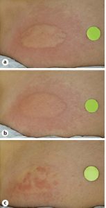
The most common adverse effect from cryotherapy is cold urticaria commonly known as cold induced hives. This reaction is marked by smooth, slightly elevated patches, which will have a red or white appearance in lighter skin tones.
A.  |
B. 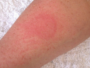 |
| Figure 5: Images A and B depict an individual with Fitzpatrick Skin Type II with ice applied. Picture B depicts the skin 5 minutes after the ice was removed. | |
In individuals with darker skin tones, cold urticaria presents as patches that are more pale than their natural skin tone. The raised patches may also present with an underlying dark brown color.
4. Iontophoresis
Iontophoresis is an intervention where electrical current is used to deliver a medication from an electrode through the skin into the subdermal tissues. One electrode has normal saline and the active electrode contains medication. A typical medication used in physical therapy is dexamethasone which is used to decrease inflammation. The most common adverse effect from iontophoresis is local skin irritation under the electrodes which can present as edema and erythema due to a histamine response in the skin.3 A more significant adverse response is the presence of small blisters under the active electrode. Skin redness on a lighter skin tone patient will be seen as “red” however on an individual with a darker skin tone this may present as a darker brown rather than red. In a person with a darker skin tone the wheal or raised patch from the histamine response maybe more noticeable than a skin color change.
Another adverse event of iontophoresis is dermatitis, which commonly presents as dryness and even cracking of the skin.
5. Tape
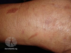
Negative skin reactions to tape can be due to mechanical trauma from removal of the tape or due to an allergic reaction to the adhesive that causes contact dermatitis. Contact dermatitis presents with erythema and edema. In patients with lighter skin tones, this looks like red, raised patches of skin. In patients with darker skin tones the patches of skin are still raised but they may present as a dark purple color or a shade darker than their skin tone. Contact dermatitis may also present with oozing, crusty skin and blisters.4
 |
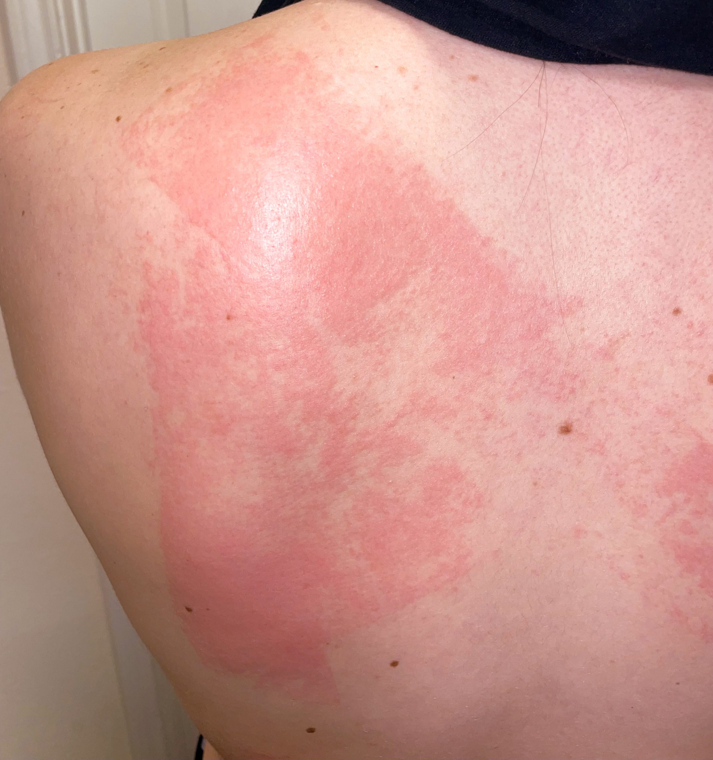 |
| Image 6: The images above show contact dermatitis after being taped with Leukotape. This depicts an individual with a lighter skin tone, Fitzpatrick skin type II. | |
References
- Cheatham SW, Baker R, Kreiswirth E. Instrument assisted soft-tissue mobilization: a commentary on clinical practice guidelines for rehabilitation professionals. Int J Sports Phys Ther. 2019;14(4): 670-682.
- Cameron MH. Physical agents in rehabilitation: an evidence-based approach to practice. 6th edition. 2023. ISBN: 9780323761949
- Falcone D, Uzunbajakava, Richters R, van de kerkhof PCM, van Erp PEJ. Histamine iontophoresis as in vivo model to study human skin inflammation with minimal barrier impairment: pilot study results of application of the model to a sensitive skin panel. Skin Pharmacology and Physiology. 2017; 30(5): 246-259.
- Li Y, Li L. Contact Dermatitis: Classifications and Management. Clin Rev Allergy Immunol. 2021;61(3):245-281. doi:10.1007/s12016-021-08875-0
