
46 Cardiac pacemaker cells
Learning Objectives
After studying this section, you should be able to-
- List the phases of cardiac autorhythmic cell action potentials and explain the ion movements that occur in each phase.
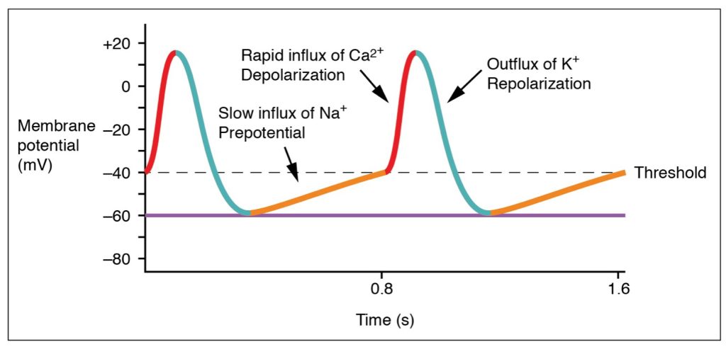
Action potentials are considerably different between cardiac pacemaker cells and cardiac muscle cells. While Na+ and K+ play essential roles, Ca2+ is also critical for both types of cells. Unlike skeletal muscles and neurons, cardiac pacemaker cells do not have a stable resting potential. Pacemaker cells contain a series of Na+ channels that allow a normal and slow influx of Na+ ions that causes the membrane potential to rise slowly from an initial value of −60 mV up to about –40 mV. This is called drift. The resulting movement of Na+ ions creates a spontaneous depolarization and brings the cell to threshold. At this point voltage-gated Ca2+ channels open and Ca2+ influx rapidly causes depolarization until it reaches a value of approximately +5 mV. At this voltage, the Ca2+ channels close and voltage-gated K+ channels open, allowing K+ efflux which causes repolarization. When the membrane potential reaches approximately −60 mV, the K+ channels close and voltage-gated slow Na+ channels open, and the drift phase begins again. This phenomenon explains the autorhythmicity properties of cardiac muscle (Figure 45.1).
Adapted from Anatomy & Physiology by Lindsay M. Biga et al, shared under a Creative Commons Attribution-ShareAlike 4.0 International License, chapter 19
cells within the heart that spontaneously depolarize at an intrinsic rate control the speed at which the heart pumps blood
