
7 Cell membrane
Learning Objectives
After reading this section, you should be able to-
- Describe the chemical composition, general structure (i.e., fluid mosaic model), and properties of all cellular membranes.
- Describe the structure of the plasma (cell) membrane, including its composition and arrangement of lipids, proteins, and carbohydrates.
- Describe the functions of different plasma membrane proteins (e.g., structural proteins, receptor proteins, channels).
Despite differences in structure and function, all living cells in multicellular organisms have a surrounding cell membrane. Just as the outer layer of your skin separates your body from its environment, the cell membrane (also known as the plasma membrane) separates the inner contents of a cell from its exterior environment. This membrane provides a protective barrier around the cell and regulates which materials can pass in or out.
Structure and Composition of the Cell Membrane
The cell membrane is an extremely pliable structure composed primarily of two layers of phospholipids (a “bilayer”). Cholesterol and various proteins are also embedded within the membrane, giving it a variety of functions, such as the ones described below.
A single phospholipid molecule has a phosphate group on one end, called the “head,” and two side-by-side chains of fatty acids that make up the lipid “tails” (Figure 7.1). The lipid tails of one layer face the lipid tails of the other layer, meeting at the interface of the two layers. The phosphate heads face outward, one layer exposed to the interior of the cell and one layer exposed to the exterior (Figure 7.1).
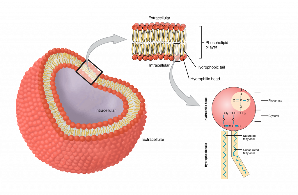
The phosphate group is negatively charged, making the head polar and hydrophilic—or “water loving.” A hydrophilic molecule is one that is attracted to water. The phosphate heads are thus attracted to the water molecules of both the extracellular and intracellular environments. The lipid tails, on the other hand, are uncharged, or nonpolar, and are hydrophobic—or “water fearing.” A hydrophobic molecule (or region of a molecule) repels and is repelled by water.
Since the phosphate groups are polar and hydrophilic, they are attracted to water in the intracellular fluid. Intracellular fluid (ICF) is the fluid inside the cell. The phosphate groups are also attracted to the extracellular fluid. Extracellular fluid (ECF) is the fluid environment outside the enclosure of the cell membrane (see above Figure). Since the lipid tails are hydrophobic, they meet in the inner region of the membrane, excluding watery intracellular and extracellular fluid from this space. In addition to phospholipids and cholesterol, the cell membrane has many proteins detailed in the next section.
Membrane Proteins
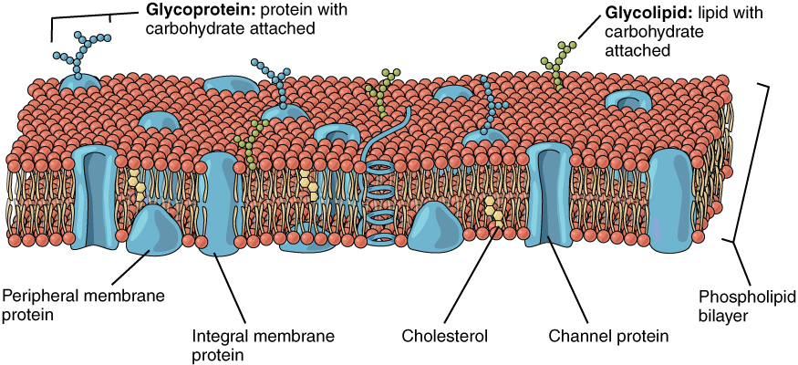
The lipid bilayer forms the basis of the cell membrane, but it is peppered throughout with various proteins. Two different types of proteins that are commonly associated with the cell membrane are integral and peripheral proteins (Figure 7.2). As its name suggests, an integral protein is a protein that is embedded in the membrane. Many different types of integral proteins exist, each with different functions. For example, an integral protein that extends an opening through the membrane for ions to enter or exit the cell is known as a channel protein. Peripheral proteins are typically found on the inner or outer surface of the lipid bilayer but can also be attached to the internal or external surface of an integral protein.
Some integral proteins serve as cell recognition proteins, or surface identity proteins, which mark a cell’s identity so that it can be recognized by other cells. Other integral proteins act as enzymes, or in cell adhesion, between neighboring cells. A receptor is a type of recognition protein that can selectively bind a specific molecule outside the cell, and this binding induces a chemical reaction within the cell. Some integral proteins serve dual roles as both a receptor and an ion channel. One example of a receptor-channel interaction is the receptors on nerve cells that bind neurotransmitters, such as dopamine. When a dopamine molecule binds to a dopamine receptor protein, a channel within the transmembrane protein opens to allow certain ions to flow into the cell. Peripheral proteins are often associated with integral proteins along the inner cell membrane where they play a role in cell signaling or anchoring to internal cellular components (i.e., cytoskeleton).
Adapted from Anatomy & Physiology by Lindsay M. Biga et al, shared under a Creative Commons Attribution-ShareAlike 4.0 International License, chapter 3.
also known as the plasma membrane; a selectively permeable barrier separating the environment inside of a cell from the outside environment
a substance that is attracted to water
a substance that repels water
fluid contained within cells
body fluids located outside of cells
proteins embedded within the cell membrane that assist with the movement of molecules into and out of the cell
Proteins found on the inner and outer surfaces of cells that assist with support and communication
a type of integral protein extending into the extracellular space, identifying the cell as self or non-self
proteins that speed up (catalyze) chemical reactions
a protein that senses specific molecules
