
27 Olfaction
Learning Objectives
After reading this section, you should be able to-
- Explain the process by which odorants activate olfactory receptors.
- Trace the path of olfaction from the olfactory receptors, to the initiation of an action potential in the olfactory nerves, through the olfactory bulb, the olfactory tract, and to the various parts of the brain.
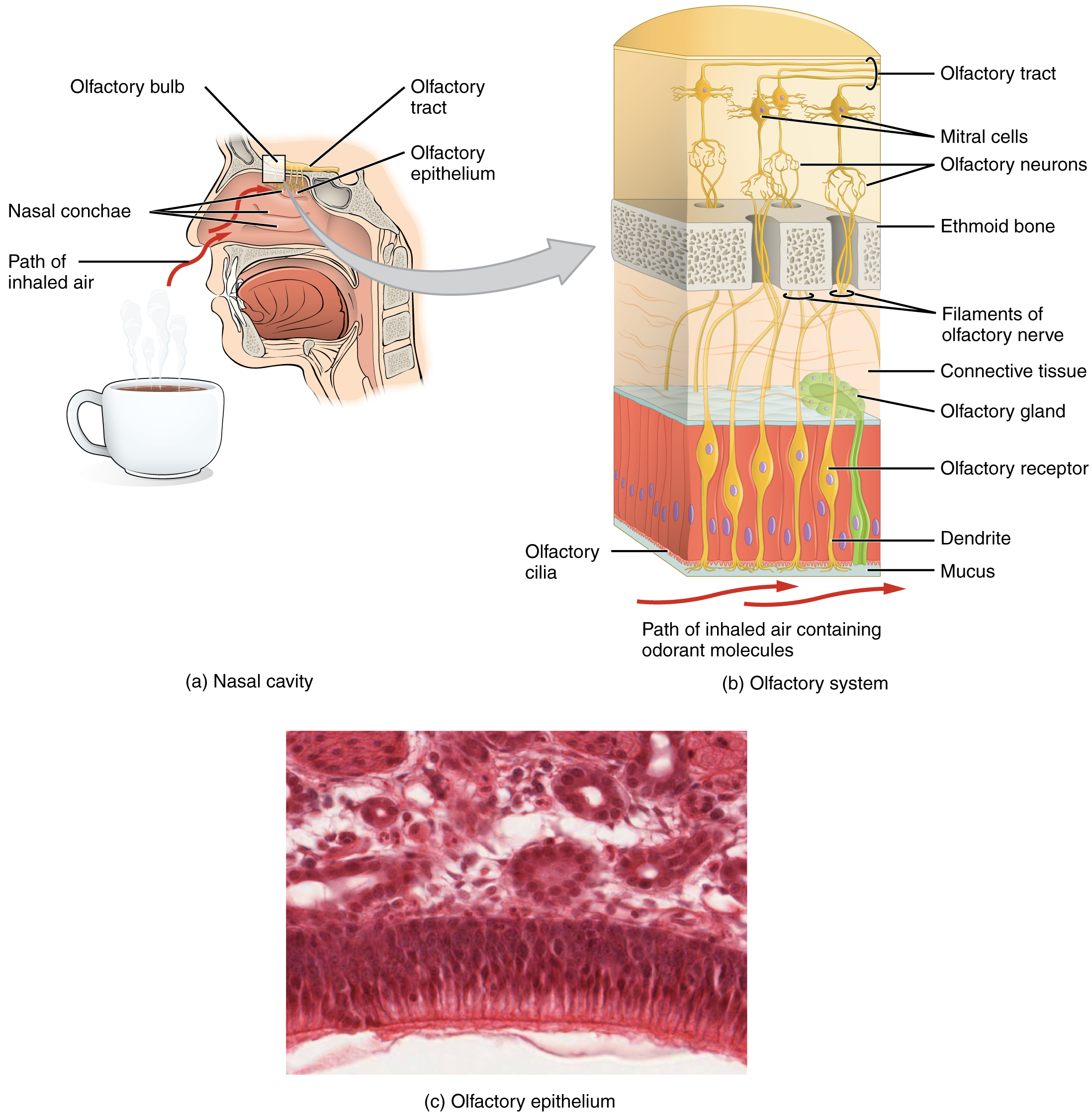
Like taste, the sense of smell, or olfaction, is also responsive to chemical stimuli. The olfactory receptor neurons are located in a small region within the superior nasal cavity (Figure 27.1). This region is referred to as the olfactory epithelium and contains bipolar sensory neurons. Each olfactory sensory neuron has dendrites that extend from the apical surface of the epithelium into the mucus lining the cavity. As airborne molecules are inhaled through the nose, they pass over the olfactory epithelial region and dissolve into the mucus. These odorant molecules bind to odarant binding proteins that keep them dissolved in the mucus and help transport them to the olfactory dendrites. The odorant–protein complex binds to a receptor protein within the cell membrane of an olfactory dendrite. These receptors are G protein–coupled, and will produce a graded membrane potential in the olfactory neurons.
When an odorant molecule binds to its corresponding receptor, it triggers a conformational change in the receptor protein, activating the associated G-protein. The activated G-protein stimulates the enzyme adenylate cyclase, leading to the production of cyclic AMP (cAMP) from ATP. Cyclic AMP acts as a second messenger by directly opening cyclic nucleotide-gated (CNG) channels, leading to the influx of calcium and sodium ions into the olfactory sensory neuron. This ion influx generates an action potential that is transmitted along the olfactory nerve to the olfactory bulb in the brain, initiating the olfactory perception of the detected odor.
The axon of an olfactory neuron extends from the basal surface of the epithelium, through an olfactory foramen in the cribriform plate of the ethmoid bone, and into the brain. The group of axons called the olfactory tract connect to the olfactory bulb on the ventral surface of the frontal lobe. Within the olfactory bulb, the axons converge into structures called glomeruli, where they synapse with mitral and tufted cells. The olfactory tract then carries the signal to several brain regions Some travel to the cerebrum, specifically to the primary olfactory cortex that is located in the inferior and medial areas of the temporal lobe. Others project to structures within the limbic system and hypothalamus, where smells become associated with long-term memory and emotional responses. This is how certain smells trigger emotional memories, such as the smell of food associated with one’s birthplace. Smell is the one sensory modality that does not synapse in the thalamus before connecting to the cerebral cortex. As a result, scents can not wake us from sleep: they do not excite the reticular activating system. This is why we use smoke detectors to alert us to fire danger using sound and smelling salts that burn the nasal epithelium to wake unconscious individuals. However, the intimate connection between the olfactory system and the cerebral cortex is one reason why smell can be a potent trigger of memories and emotion.
The nasal epithelium, including the olfactory cells, can be harmed by airborne toxic chemicals. Therefore, the olfactory neurons are regularly replaced within the nasal epithelium, after which the axons of the new neurons must find their appropriate connections in the olfactory bulb. These new axons grow along the axons that are already in place in the cranial nerve, ensuring proper connectivity and functionality.
Adapted from Anatomy & Physiology by Lindsay M. Biga et al, shared under a Creative Commons Attribution-ShareAlike 4.0 International License, chapter 15.
the sensation of smell
the region of the nasal cavity that contains bipolar neurons responsible for olfaction
bipolar neurons within the olfactory epithelium that transmit smell sensations to the brain
specialized proteins with the ability to bind guanosine triphosphate (GTP) and guanosine diphosphate (GDP)
ion channels that respond to cyclic nucleotides, such as cAMP and cGMP
the neural structure that receives input about smells detected by the bipolar olfactory neurons in the olfactory epithelium
