
14 Graded potentials
Learning Objectives
After reading this section, you should be able to-
- Define and describe depolarization, repolarization, hyperpolarization, and threshold.
- Define excitatory postsynaptic potential (EPSP) and inhibitory postsynaptic potential (IPSP) and interpret graphs showing the voltage vs. time relationship of an EPSP and an IPSP
- Explain temporal and spatial summation of synaptic potentials.
- Explain how a single neurotransmitter may be excitatory at one synapse and inhibitory at another
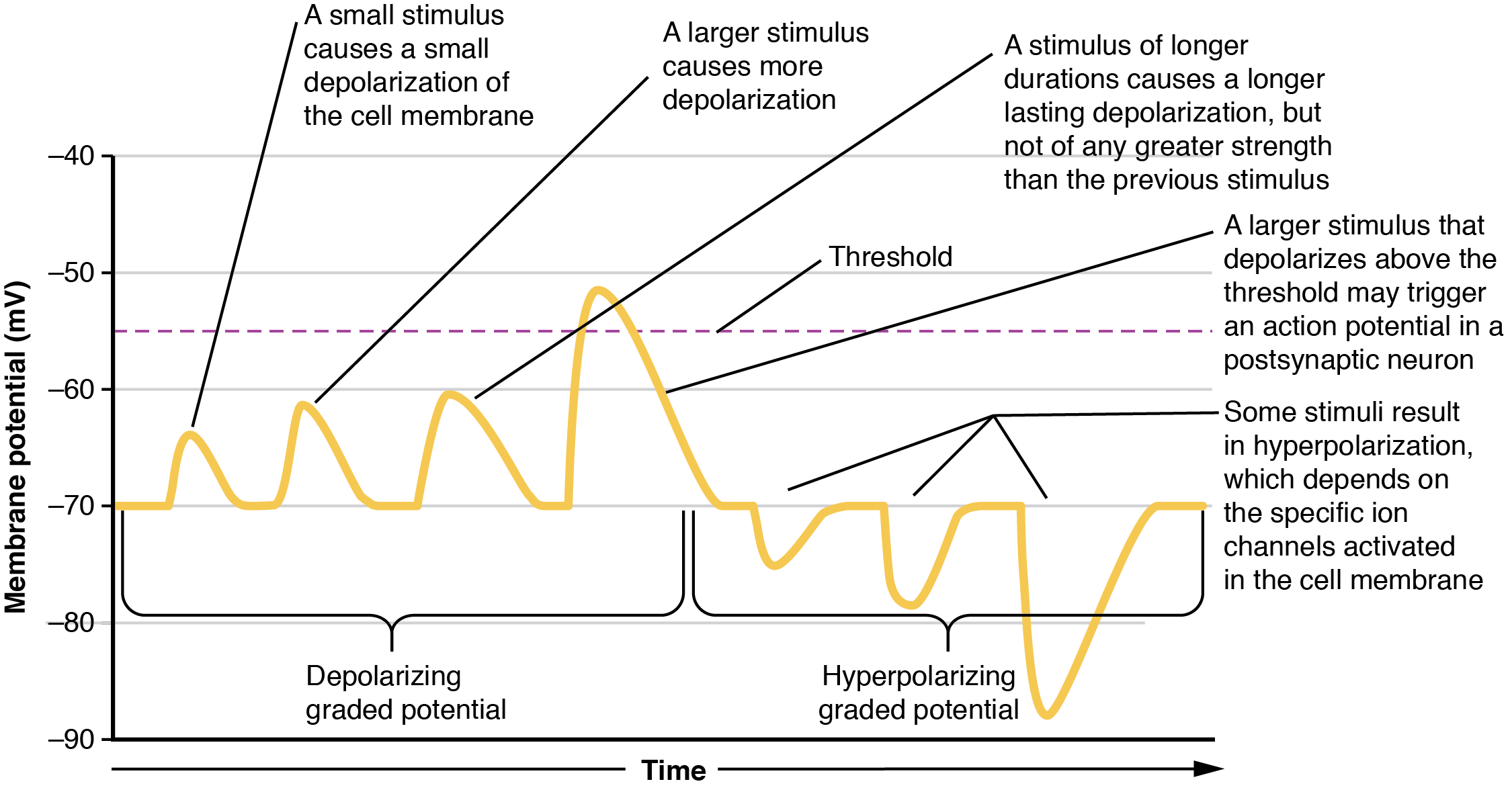
Local changes in the membrane potential away from resting levels are called graded potentials, sometimes referred to as a local potential, and are usually associated with the opening gated channels on the membrane a neuron. The type and amount of change in the membrane potential is determined by the specific ion that crosses the membrane, how many ions cross and for how long. Graded potentials can be of two sorts, either they are depolarizing (above resting membrane potential) or hyperpolarizing (below resting membrane potential) (Figure 14.1).
Depolarizing graded potentials are often the result of Na+ or Ca2+ entering the cell. Both of these ions are more highly concentrated outside of the cell than inside; because they have a positive charge, the cell membrane becomes less negative inside the cell relative to the outside when these ions move into the cell. An example of a depolarizaing graded potential could take membrane potential from –70 mV → –60 mV. This would be a 10 mV depolarizing graded potential.
Hyperpolarizing graded potentials can be caused by K+ leaving the cell or Cl– entering the cell. The membrane becomes more negative if a positive charge moves out of a cell or if a negative charge enters the cell. Graded potentials are transient and dissipate as they move away from the site of the initial stimulus. An example of a hyperpolarizing graded potential could take membrane potential from –70 mV → –80 mV. This would be a 10 mV hyperpolarizing graded potential.
When ion channels are left open longer or when more channels are opened (for the same ion), the stimulus affecting a neuron is bigger. A “bigger stimulus” occurs due to a more painful stimulus, a heavier load, a brighter light etc. These larger stimuli induce larger graded potentials of longer duration in neurons and can be either depolarizing or hyperpolarizing.
For the unipolar cells of sensory neurons—both those with free nerve endings and those within encapsulations—graded potentials develop in the dendrites and influence the generation of an action potential in the axon of the same cell. This is called a generator potential. For other sensory receptor cells which are not neurons, such as taste cells or photoreceptors of the retina, graded potentials in receptor cell membranes result in the release of neurotransmitters at synapses with sensory neurons. This is called a receptor potential, and we will consider this type of graded potential during a discussion of the special senses.
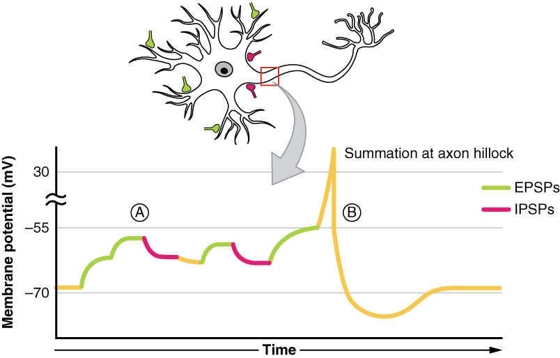
A postsynaptic potential (PSP) is the graded potential in the dendrites or cell body of a neuron that is receiving synapses from other cells. Postsynaptic potentials can be depolarizing or hyperpolarizing. Depolarization in a postsynaptic potential is called an excitatory postsynaptic potential (EPSP) because it causes the membrane potential to move toward threshold. Hyperpolarization in a postsynaptic potential is called an inhibitory postsynaptic potential (IPSP) because it causes the membrane potential to move away from threshold.
Mechanism at a glance:
-
EPSP: Opening Na⁺ (or Ca²⁺) channels → inward positive current → depolarization toward ~0 mV
-
IPSP: Opening Cl⁻ (or K⁺) channels → inward negative/outward positive current → hyperpolarization away from threshold
Example
A neurotransmitter-gated Na⁺ conductance opens briefly → Vm goes from –70 mV to –60 mV (depolarization of +10 mV). When it closes, passive K⁺ efflux returns Vm to –70 mV (repolarization), and a larger-than-normal K⁺ conductance may overshoot to –75 mV (hyperpolarization).
Summation
All types of graded potentials will result in small changes (either depolarizing or hyperpolarizing) in the voltage of a membrane. These changes can lead to the neuron reaching threshold if the changes add together, or summate. The combined effects of different types of graded potentials are illustrated in Figure 14.2. If the total change in voltage that reaches the initial segment (or trigger zone) is a positive 15 mV, meaning that the membrane depolarizes from -70 mV (resting membrane potential) to -55 mV (threshold), then the graded potentials will result in the initiation of an action potential.
Graded potentials summate at a specific location at the beginning of the axon to initiate the action potential, namely the initial segment. For sensory neurons, the initial segment is directly adjacent to the dendritic endings (since the cell body is located more proximally). For all other neurons, the initial segment of the axon is found at the axon hillock and it is where summation takes place. These locations have a high density of voltage-gated Na+ channels that initiate the depolarizing phase of the action potential and is often referred as the trigger zone.
Summation can be spatial or temporal, meaning it can be the result of multiple graded potentials occurring simultaneously at different locations on the neuron (spatial), or all at the same place but in rapid succession (temporal). Spatial and temporal summation can act together, as well. Since graded potentials dissipated with distance and time, summation is the total change in voltage due to all spatial and temporal graded potentials that reach the trigger zone or initial segment at each moment. When these local potentials sum to reach threshold at the axon hillock, an all-or-none action potential is triggered
Adapted from Anatomy & Physiology by Lindsay M. Biga et al, shared under a Creative Commons Attribution-ShareAlike 4.0 International License, chapter 12.
small, short lasting changes in membrane potential
a difference in membrane potential produced by the activation of a sensory receptor
a potential that leads to the release of a neurotransmitter at a synapse with a sensory neuron
changes in membrane potential that move the cell away from its resting state
a potential that causes the cell membrane to depolarize and move closer to threshold
a potential that causes the cell membrane to hyperpolarize and move further away from threshold
the ability of a membrane to reach threshold due to the addition of multiple graded potentials
summation occurring due to multiple graded potentials occurring simultaneously at different locations on a neuron
summation occurring due to multiple graded potentials occurring simultaneously in rapid succession at the same place on a neuron
