
39 Whole muscle contraction
Learning Objectives
After reading this section, you should be able to-
- Interpret a graph of the length-tension relationship and describe the anatomical basis for that relationship.
- Compare and contrast isotonic and isometric contraction.
- Compare and contrast concentric and eccentric contraction.
To move an object, referred to as a load, the muscle fibers of a skeletal muscle must shorten. The force generated by a contracting muscle is called muscle tension. Muscle tension can also be generated when the muscle is contracting against a load that does not move, resulting in two main types of skeletal muscle contractions: isotonic contractions and isometric contractions (Figure 39.1).
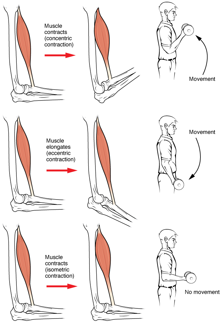
In isotonic contractions, where the tension in the muscle stays relatively constant, a load is moved as the length of the muscle changes. A concentric contraction involves the muscle producing tension and shortening to move a load. An example of this is the contraction of the biceps brachii muscle when a hand weight is brought upward toward the body. An eccentric contraction occurs when the muscle tension produced is less than the load and a muscle lengthens while under tension. This type of contraction is observed when the same hand weight is lowered in a slow and controlled manner by the biceps brachii. Both concentric and eccentric contractions involve force production by the muscle and cross-bridge cycling with the myosin heads pulling toward the M-line. The only difference between the two is whether the muscle length is shortening or elongating during the contraction.
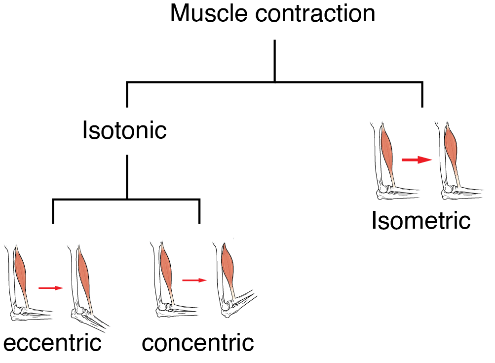
An isometric contraction occurs when a muscle produces tension without a change in muscle length. Isometric contractions involve sarcomere shortening and increasing muscle tension. However, a load is not moved, as the force produced cannot overcome the resistance provided by the load. For example, if one attempts to lift a hand weight that is too heavy, there will be sarcomere activation and shortening to a point, and ever-increasing muscle tension, but no change in the position of the hand weight. In everyday living, isometric contractions are active in maintaining posture and maintaining bone and joint stability.
Most actions of the body are the result of a combination of isotonic and isometric contractions working together to produce a wide range of outcomes. These muscle activities are under the control of the nervous system. A crucial aspect of nervous system control of skeletal muscles is the role of motor units.
Motor Units
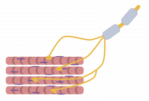
As previously discussed, the contraction of skeletal muscle fibers is triggered by signaling from a motor neuron. Each muscle fiber is innervated by only one motor neuron, but a single motor neuron can innervate multiple muscle fibers. A motor unit is defined as a single motor neuron and all of the muscle fibers it innervates (Figure 39.3). The size of a motor unit dictates its function. A small motor unit, composed of a motor neuron and only a few muscle fibers, permits very fine motor control of a muscle. For example, the extraocular eye muscles have thousands of muscle fibers with every 5 – 10 fibers supplied by a single motor neuron; this allows for exquisite control of eye movements so that both eyes can quickly focus on an object. Small motor units are also involved in the many fine movements of the fingers and thumb of the hand for grasping, texting, etc.
Large motor units have more muscle fibers per neuron than small motor units. Larger motor units are concerned with simple, or “gross,” movements, such as moving parts of the body against gravity. The large motor units of the thigh muscles or back muscles, where a single motor neuron will supply thousands of muscle fibers in a muscle, are representative of this type of activity.
Most muscles in the human body have a mixture of small and large motor units which gives the nervous system a wide range of control over the muscle. The smaller motor units in a muscle have motor neurons that are more excitable. Initial activation of these smaller motor units results in a relatively small degree of tension generated in a muscle. As more strength is needed, larger motor units are enlisted to generate more tension. This process of bringing on additional motor units to produce more tension is known as recruitment. This process allows a muscle such as the biceps brachii to pick up a feather with minimal force generation versus picking up a heavy weight, which requires a much greater amount of force generation.
When necessary, the maximal number of motor units in a muscle can be recruited simultaneously, producing the maximum force of contraction for that muscle, but this cannot last for very long because of the energy requirements to sustain the contraction. To prevent complete muscle fatigue, motor units are generally not all simultaneously active, but instead some motor units rest while others are active, which allows for longer muscle contractions. The nervous system thus uses recruitment as a mechanism to efficiently utilize a skeletal muscle.
The Length-Tension Range of a Sarcomere
As discussed previously, when a skeletal muscle fiber contracts, myosin heads attach to actin to form cross-bridges, followed by the thin filaments sliding over the thick filaments as the heads pull the actin. This results in sarcomere shortening, creating the tension of the muscle contraction. The cross-bridges can only form where thin and thick filaments overlap; thus, the length of the sarcomere has a direct influence on the force generated when the sarcomere shortens. This is called the length-tension relationship.
The ideal length of a sarcomere to produce maximal tension occurs at 80 percent to 120 percent of its resting length, with 100 percent being the state where the medial edges of the thin filaments are just at the most-medial myosin heads of the thick filaments (Figure 39.4). This length maximizes the overlap of actin-binding sites and myosin heads.
If a sarcomere is stretched past the ideal length (beyond 120 percent), thick and thin filaments do not fully overlap, which results in less tension produced. If the muscle is stretched to the point where the thick and thin filaments do not overlap at all, no cross-bridges can be formed, and no tension is generated. This amount of stretching does not usually occur as accessory proteins and connective tissue oppose extreme stretching.

If a sarcomere is shortened beyond 80 percent, the zone of overlap is reduced with the thin filaments jutting beyond the last of the myosin heads. Eventually, there is nowhere else for the thin filaments to go and the amount of tension is diminished.
Adapted from Anatomy & Physiology by Lindsay M. Biga et al, shared under a Creative Commons Attribution-ShareAlike 4.0 International License, chapter 10.
force generated by a contracting muscle
a muscle contraction defined by constant tension of a muscle, while it changes length to move a load
an isotonic contraction defined by the length of a muscle shortening as it produces constant tension to move a load
an isotonic contraction defined by the lengthening of a muscle as it produces less tension than the load
the repetitive processes of myosin heads binding to actin, pulling to form cross-bridges, detaching, and re-cocking
a muscle contraction defined by the production of tension without a change in muscle length
a single motor neuron and all of the muscle fibers it innervates
the activation of additional motor units to produce a given muscle contraction
