29 Functions of the endocrine system
Learning Objectives
After reading this section you should be able to-
- Describe the major functions of the endocrine system
- Define the terms hormone, endocrine gland, endocrine tissue, and target cell
- Compare and contrast how the nervous and endocrine systems control body function, with emphasis on the mechanisms by which the controlling signals are transferred through the body and the time course of the response(s) and action(s).
- List and describe several types of stimuli that control production and secretion of hormones
- Describe the roles of negative and positive feedback in controlling hormone release
Communication within the human body involves the transmission of signals to control and coordinate actions in an effort to maintain homeostasis. There are two major organ systems responsible for providing these communication pathways: the nervous system and the endocrine system. The nervous system is primarily responsible for rapid communication throughout the body. As discussed in previous chapters, the nervous system utilizes two types of signals – electrical and chemical (Table 29.1). Electrical signals are sent via the generation and propagation of action potentials which move along the membrane of a cell. Once the action potential reaches the synaptic terminal, the electrical signal is converted to a chemical signal as neurotransmitters are released into the synaptic cleft. When the neurotransmitters bind with receptors on the receiving (post-synaptic) cell, a new electrical signal is generated and quickly continues on to its destination. In this way, neural communication enables body functions that involve quick, brief actions, such as movement, sensation, and cognition.
Stimuli Controlling Hormone Production
The production and secretion of hormones by endocrine glands are tightly regulated by various stimuli, including changes in the internal and external environment, physiological needs, and feedback mechanisms. These stimuli serve as signals that prompt endocrine glands to release specific hormones in response to specific physiological requirements.
One example of a stimulus controlling hormone production is the regulation of insulin secretion by blood glucose levels. When blood glucose levels rise, such as after a meal, pancreatic beta cells sense this increase and respond by secreting insulin into the bloodstream. Insulin acts to facilitate the uptake of glucose by cells, thereby reducing blood glucose levels and restoring homeostasis. Conversely, when blood glucose levels decrease, insulin secretion decreases, allowing for the release of glucose from storage sites to maintain glucose homeostasis.
Similarly, the hypothalamus-pituitary-adrenal (HPA) axis responds to stress-related stimuli by releasing adrenocorticotropic hormone (ACTH), which stimulates the adrenal glands to produce cortisol. During periods of stress, such as physical injury or psychological distress, the hypothalamus detects the stressor and releases corticotropin-releasing hormone (CRH), which signals the pituitary gland to release ACTH. In turn, ACTH stimulates the adrenal cortex to secrete cortisol, initiating the body’s stress response and helping to mobilize energy reserves to cope with the stressor.
Other stimuli controlling hormone production include changes in blood levels of ions or nutrients (humoral stimuli), hormonal signals from other endocrine glands (hormonal stimuli), and neural signals from the autonomic nervous system (neural stimuli). For example, the release of parathyroid hormone (PTH) by the parathyroid glands is stimulated by low blood calcium levels, triggering PTH secretion to increase calcium levels by promoting its release from bone and enhancing its reabsorption from the kidneys.
Moreover, hormonal signals from other endocrine glands, such as thyroid-stimulating hormone (TSH) from the anterior pituitary, regulate the production and secretion of hormones from target glands like the thyroid gland. TSH stimulates the thyroid gland to produce thyroid hormones (T3 and T4), which regulate metabolism and energy balance.
Feedback Mechanisms in Hormone Release
- Negative Feedback: Most hormone regulation operates via negative feedback, where an increase in a hormone’s effect inhibits further hormone release. For example, high blood glucose levels stimulate insulin release, which lowers glucose levels. As glucose levels decrease, insulin release is reduced, maintaining homeostasis.
- Positive Feedback: In positive feedback, the effect of a hormone causes more of the hormone to be released. An example is oxytocin during childbirth. Oxytocin release causes uterine contractions, which stimulate more oxytocin release, intensifying the contractions until delivery.
Endocrine Organs
Hormones are released by secretory cells that are derived from epithelial tissue. Often, these cells are clustered together, forming endocrine glands. Unlike exocrine glands, which have a duct for conveying secretions to the outside of the body (e.g., sweat gland), endocrine glands secrete substances directly into the surrounding interstitial fluid. From there, hormones enter the bloodstream for distribution throughout the body.
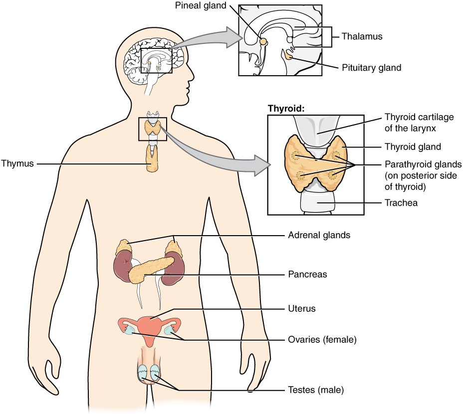
The major endocrine glands found in the human body include the pituitary gland, thyroid gland, parathyroid glands, thymus gland, adrenal glands, pineal gland, testes, and ovaries (Figure 29.1). While some of the glands are purely endocrine (e.g., thyroid gland), others serve both endocrine and exocrine function. For example, the pancreas contains cells that secrete digestive enzymes and juices into the small intestine (exocrine function) and cells that secrete the hormones insulin and glucagon, which regulate blood glucose levels.
In addition to the endocrine glands, major organs of the body show endocrine function including the hypothalamus, heart, kidneys, stomach, small intestine, and liver. In these organs, the tissues that release hormones are referred to as endocrine tissue. Moreover, adipose tissue has long been known to produce hormones, and recent research has revealed that bone tissue plays a role in hormone production and secretion.

Adapted from Anatomy & Physiology by Lindsay M. Biga et al, shared under a Creative Commons Attribution-ShareAlike 4.0 International License, chapter 17.
the space between the axon of one neuron and the dendrites of another neurons; the site where electrical signals are transmitted from one neuron to the next
the messenger system that utilizes chemicals to send signals into the circulation that target and regulate other tissues and organs
chemical messengers released by endocrine cells that regulate other cells within the body
neuroendocrine system responsible for regulating the stress response
changes in extracellular fluids that trigger the release of hormones
release of one hormone in response to the release of another hormone
autonomic signals that result in the release of hormones from endocrine glands
an organ that produces or secretes hormones that are released into the bloodstream

