
18 Synapse
Learning Objectives
After reading this section, you should be able to-
- Define a synapse and explain the difference between an electrical synapse and a chemical synapse.
- Describe the structures involved in a typical chemical synapse (e.g., axon terminal [synaptic knob], voltage-gated calcium channels, synaptic vesicles of presynaptic cell, synaptic cleft, neurotransmitter receptors of the postsynaptic cell).
- Describe the events of synaptic transmission in proper chronological order from the release of neurotransmitter by synaptic vesicles to the effect of the neurotransmitter on the postsynaptic cell.
- Describe the different mechanisms (e.g., reuptake, enzymatic breakdown, diffusion) by which neurotransmitter activity at a synapse can be terminated
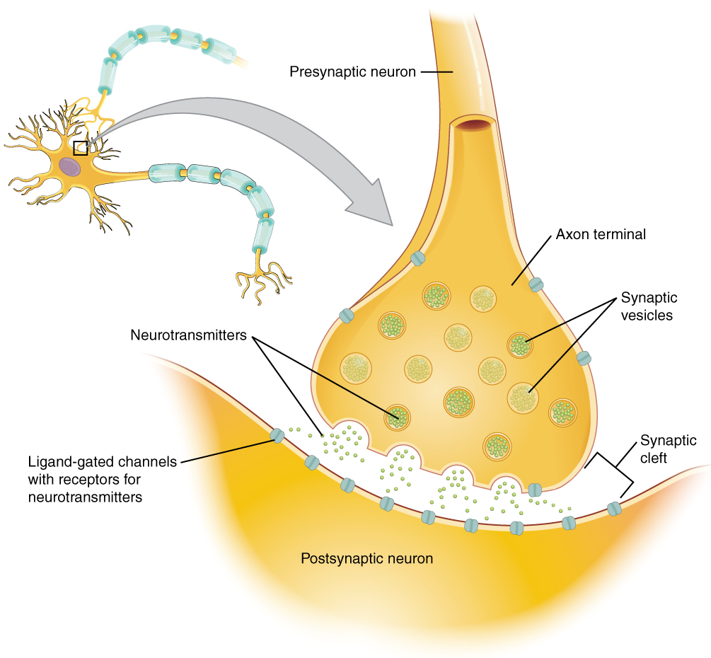
Synapses
A synapse is the site of communication between a neuron and another cell. There are two types of synapses: chemical synapses and electrical synapses. In a chemical synapse, a chemical signal, which is called a neurotransmitter, is released from the neuron and binds to a receptor on the other cell. In an electrical synapse, the membranes of two cells directly connect through a gap junction so that ions can pass directly from one cell to the next, transmitting a signal. Both types of synapses occur in the nervous system, though chemical synapses are more common.
An example of a chemical synapse is the neuromuscular junction (NMJ), which will be described in the chapter on muscle tissue. In the nervous system, there are many additional synapses that utilize the same mechanisms as the NMJ. All chemical synapses have common characteristics, which can be summarized in Table 18.1:
| Example Chemical Synapse (Table 18.1) |
|
| Common Chemical Synapse Element | Specific element in a Skeletal Muscle Neuromuscular Junction |
|---|---|
| presynaptic element | somatic motor neuron axon terminal |
| neurotransmitter (packaged in vesicles) | acetylcholine |
| synaptic cleft | space between somatic motor neuron and muscle cell membrane |
| receptor proteins | nicotinic acetylcholine (cholinergic) receptor |
| postsynaptic element | motor end plate of the sarcolemma |
| neurotransmitter elimination/re-uptake | degrading enzyme: acetylcholinesterase |
Neurotransmitter Release & the Synaptic Cleft
When an action potential reaches the axon terminals, voltage-gated Ca2+ channels in the membrane of the synaptic end bulb (axon terminal) open. Ca2+ diffuses down its concentration gradient and enters into the presynaptic neuron axon terminal (end bulb). Once Ca2+ is inside the presynaptic end bulb, it binds to proteins that trigger exocytosis—the process of vesicles fusing with the cell membrane to release neurotransmitters into the synaptic cleft. The released neurotransmitter moves into the small gap between the cells, called the synaptic cleft.
Once in the synaptic cleft, the neurotransmitter diffuses the short distance to the postsynaptic membrane and can bind to neurotransmitter receptors. Receptors are specific for the neurotransmitter, and the two fit together like a lock and key, and so a neurotransmitter will not bind to receptors for other neurotransmitters (Figure 18.1). This process converts an electrical signal in the presynaptic neuron into a chemical signal across the synapse, and then back into an electrical signal in the postsynaptic cell.
The neurotransmitter is cleared from the synapse either by enzymatic degradation, neuronal reuptake, or glial reuptake. In some cases, surrounding glial cells (like astrocytes) remove neurotransmitters from the synaptic cleft. This ensures that the signal ends in a timely manner and prevents continuous stimulation of the postsynaptic cell. Together, these steps ensure that neurons can communicate efficiently, precisely, and briefly—allowing the nervous system to coordinate rapid and complex responses.
Agonists and Antagonists
Some drugs and toxins affect synaptic transmission by mimicking or blocking neurotransmitters. A substance that activates a receptor by mimicking a neurotransmitter is called an agonist. A substance that blocks a receptor and prevents the neurotransmitter from binding is called an antagonist. For example, nicotine is an agonist at certain acetylcholine receptors, while curare is an antagonist that blocks those same receptors at the neuromuscular junction.
Adapted from Anatomy & Physiology by Lindsay M. Biga et al, shared under a Creative Commons Attribution-ShareAlike 4.0 International License, chapter 12.
A period of time when membrane potential is become less negative
a change in membrane potential that occurs due to the opening and closing of voltage-gated ion channels on the cell membrane
The site of communication between a neuron and another cell
the transmission of a nerve impulse due to the release of a neurotransmitter from one cell that travels and binds to a receptor on another cell
the transmission of a nerve impulse due a direct connection between cell membranes through a gap junction, allowing ions to pass directly from one cell to the next
the discharge of substances out of the cell and into the extracellular space by the transport of secretory vesicles
the space between the axon of one neuron and the dendrites of another neurons; the site where electrical signals are transmitted from one neuron to the next
